. 2017 Oct 25;139(42):14829-14832.
doi: 10.1021/jacs.7b06730. Epub 2017 Oct 10.

An Adaptable Phospholipid Membrane Mimetic System for Solution NMR Studies of Membrane Proteins

Affiliations

An Adaptable Phospholipid Membrane Mimetic System for Solution NMR Studies of Membrane Proteins

Chih-Ta Henry Chien et al. J Am Chem Soc. .

Abstract

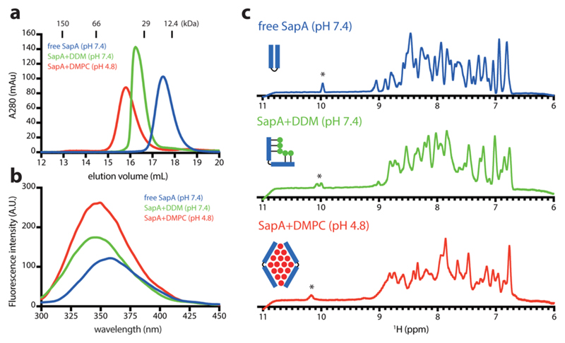
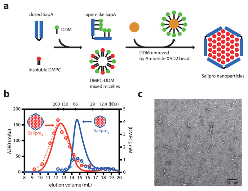
Based on the saposin-A (SapA) scaffold protein, we demonstrate the suitability of a size-adaptable phospholipid membrane-mimetic system for solution NMR studies of membrane proteins (MPs) under close-to-native conditions. The Salipro nanoparticle size can be tuned over a wide pH range by adjusting the saposin-to-lipid stoichiometry, enabling maintenance of sufficiently high amounts of phospholipid in the Salipro nanoparticle to mimic a realistic membrane environment while controlling the overall size to enable solution NMR for a range of MPs. Three representative MPs, including one G-protein-coupled receptor, were successfully incorporated into SapA-dimyristoylphosphatidylcholine nanoparticles and studied by solution NMR spectroscopy.


Conflict of interest statement

Notes

The authors declare no competing financial interest.

Figures

Figure 1
Figure 1
Biophysical characterization of free SapA, SapA-DDM, and SapA-DMPC nanoparticles. (a) Size-exclusion chromatography. (b) Fluorescence spectroscopy. (c) 1D 1H NMR spectroscopy. Tryptophan indole signals are indicated by *.
Figure 2
Figure 2
Generation of SapA-DMPC nanoparticles at pH 6. (a) Schematic representation of the SapA-DMPC nanoparticle assembly. (b) SEC of SapA-DMPC with initial mixing SapA:DMPC ratios of 1:15 (blue) and 1:45 (red) at pH 6. DMPC was quantified by molybdate assay (open circle). The proposed SapA arrangement is indicated. (c) Negative-stain EM images of Salipro3. Scale bar, 50 nm.
Figure 3
Figure 3
2D [1H,15N]-SOFAST-TROSY spectrum of non-deuterated 15N-labeled OmpX in SapA-DMPC nanoparticles.
Figure 4
Figure 4
Overlay of the 2D 13C-SOFAST-HMQC spectra of [13Cε-methionine] β1AR in the apo state (blue), the isoprenaline-bound state (green), and the isoprenaline-Nb80-bound ternary complex (red).

References

    1. Wallin E, von Heijne G. Protein Sci. 1998;7:1029. - PMC - PubMed
    1. Cross TA, Murray DT, Watts A. Eur Biophys J. 2013;42:731. - PMC - PubMed
    1. Ding Y, Fujimoto LM, Yao Y, Plano GV, Marassi FM. Biochim Biophys Acta, Biomembr. 2015;1848:712. - PMC - PubMed
    1. Zhang M, Huang R, Im SC, Waskell L, Ramamoorthy A. J Biol Chem. 2015;290:12705. - PMC - PubMed
    1. Laganowsky A, Reading E, Allison TM, Ulmschneider MB, Degiacomi MT, Baldwin AJ, Robinson CV. Nature. 2014;510:172. - PMC - PubMed

Publication types

MeSH terms

LinkOut - more resources