Review
. 2019 Feb 1;30(4):650-673.
doi: 10.1089/ars.2017.7374. Epub 2017 Nov 13.

Structural Biology of Proline Catabolic Enzymes

Affiliations
Review

Structural Biology of Proline Catabolic Enzymes

John J Tanner. Antioxid Redox Signal. .

Abstract

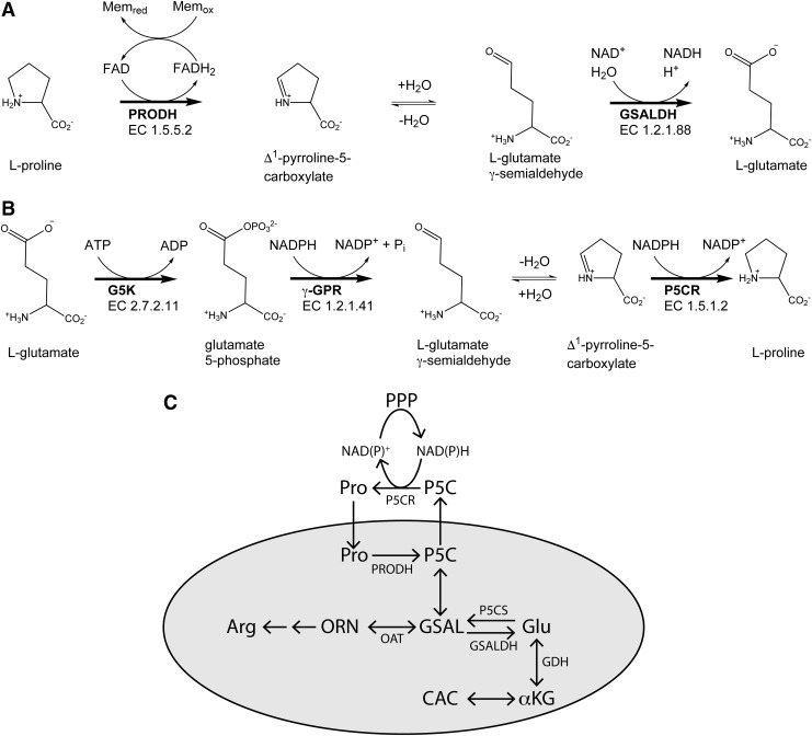
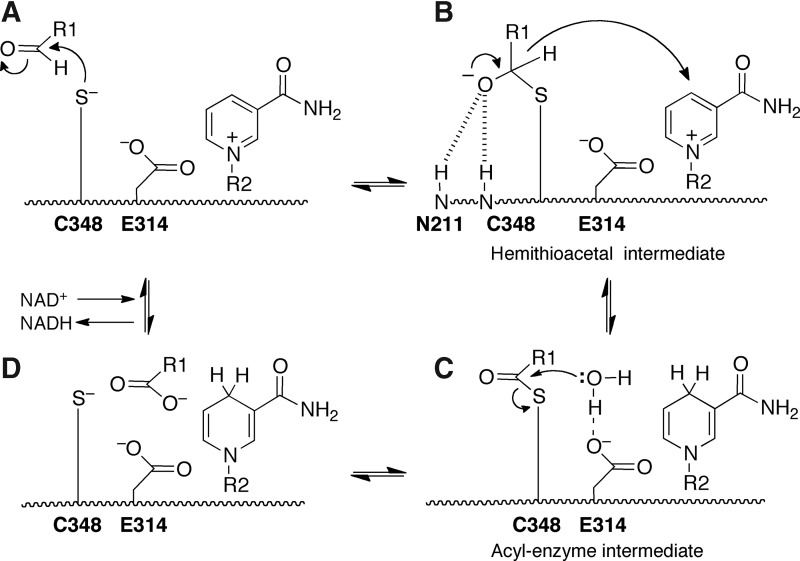
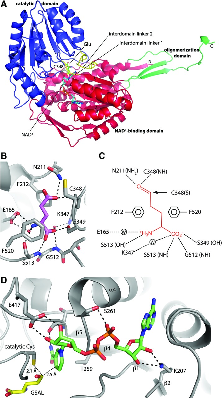
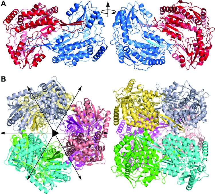
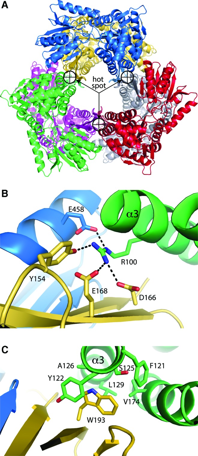
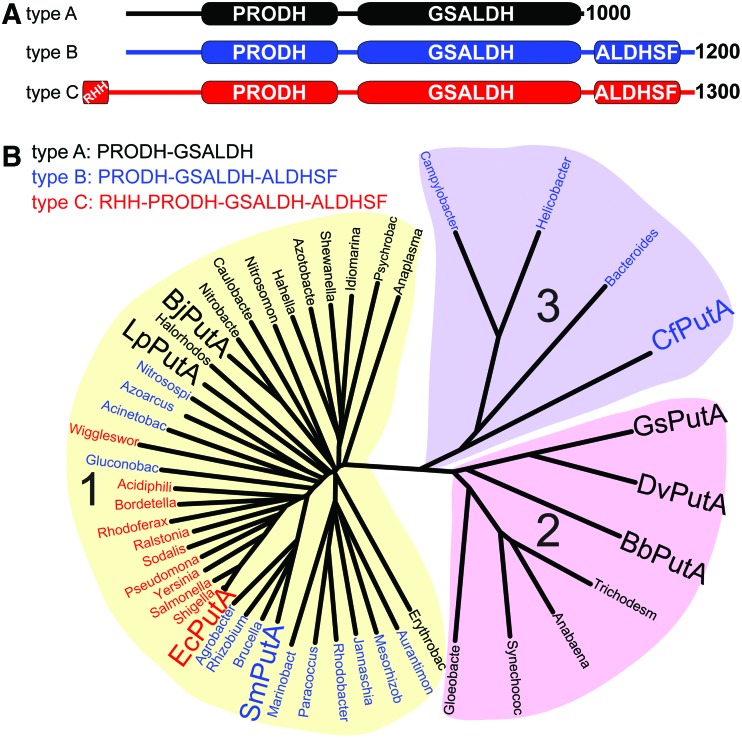
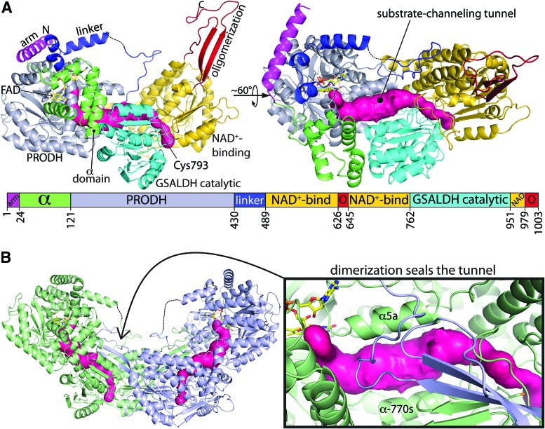
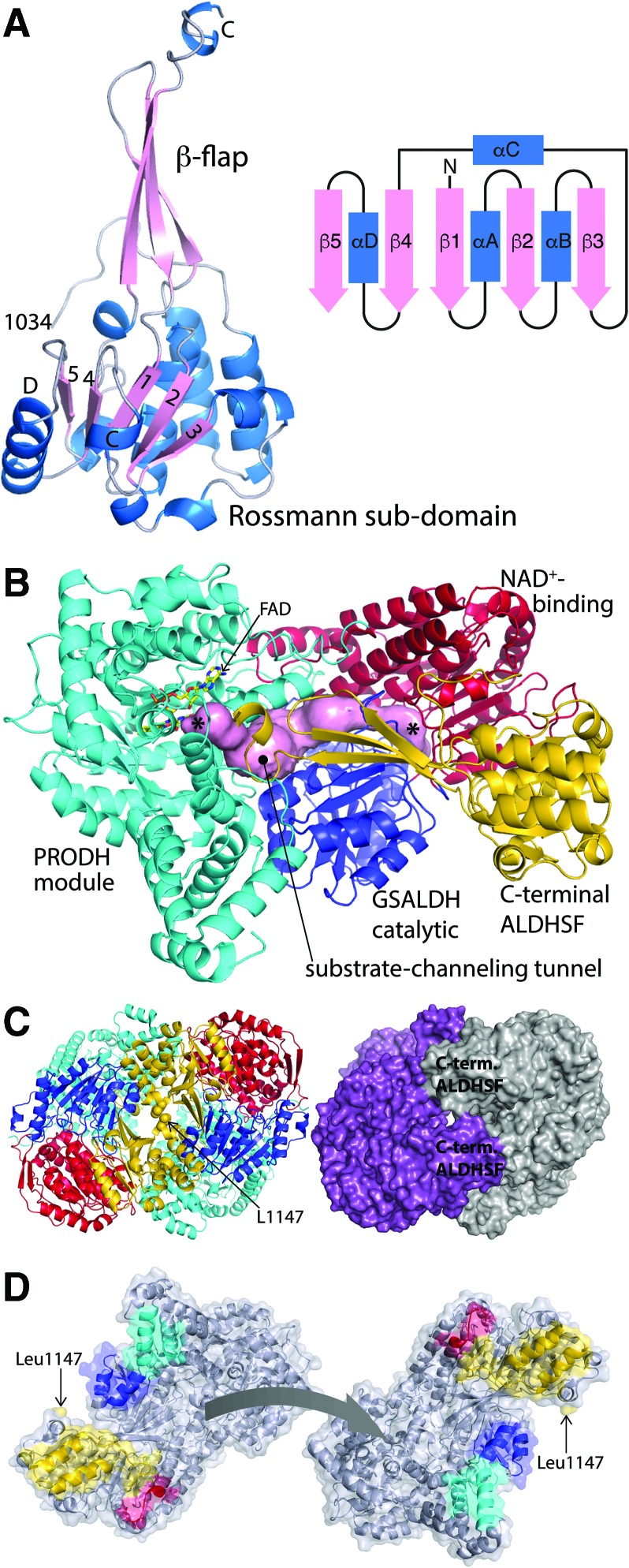
Significance: Proline catabolism refers to the 4-electron oxidation of proline to glutamate catalyzed by the enzymes proline dehydrogenase (PRODH) and l-glutamate γ-semialdehyde dehydrogenase (GSALDH, or ALDH4A1). These enzymes and the intermediate metabolites of the pathway have been implicated in tumor growth and suppression, metastasis, hyperprolinemia metabolic disorders, schizophrenia susceptibility, life span extension, and pathogen virulence and survival. In some bacteria, PRODH and GSALDH are combined into a bifunctional enzyme known as proline utilization A (PutA). PutAs are not only virulence factors in some pathogenic bacteria but also fascinating systems for studying the coordination of metabolic enzymes via substrate channeling. Recent Advances: The past decade has seen an explosion of structural data for proline catabolic enzymes. This review surveys these structures, emphasizing protein folds, substrate recognition, oligomerization, kinetic mechanisms, and substrate channeling in PutA.

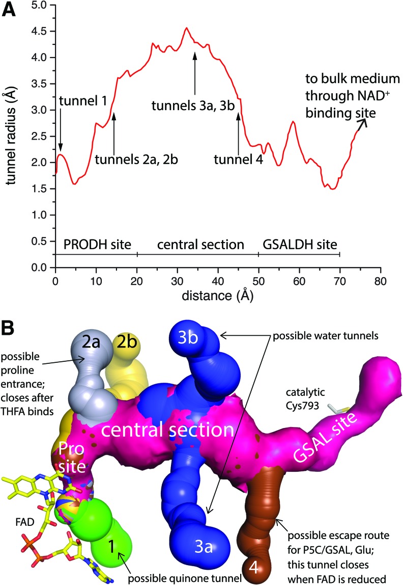
Critical issues: Major unsolved structural targets include eukaryotic PRODH, the complex between monofunctional PRODH and monofunctional GSALDH, and the largest of all PutAs, trifunctional PutA. The structural basis of PutA-membrane association is poorly understood. Fundamental aspects of substrate channeling in PutA remain unknown, such as the identity of the channeled intermediate, how the tunnel system is activated, and the roles of ancillary tunnels.

Future directions: New approaches are needed to study the molecular and in vivo mechanisms of substrate channeling. With the discovery of the proline cycle driving tumor growth and metastasis, the development of inhibitors of proline metabolic enzymes has emerged as an exciting new direction. Structural biology will be important in these endeavors.

Keywords: aldehyde dehydrogenase 4A1; proline dehydrogenase; proline utilization A; protein oligomerization; substrate channeling.


Figures

<b>FIG. 1.</b>
FIG. 1.
The reactions and enzymes of proline metabolism. (A) Proline catabolism. (B) Proline biosynthesis from glutamate. (C) Diagram depicting the subcellular locations of proline metabolic enzymes in humans and the proline cycle. The gray oval represents the mitochondria. γ-GPR, γ-glutamate phosphate reductase; G5K, glutamate 5-kinase; GSAL, l-glutamate-γ-semialdehyde; GSALDH, l-glutamate γ-semialdehyde dehydrogenase; OAT, ornithine δ-aminotransferase; ORN, ornithine; P5C, Δ1-pyrroline-5-carboxylate; P5CS, P5C synthase; PRODH, proline dehydrogenase.
<b>FIG. 2.</b>
FIG. 2.
The PRODH fold. The structure of DrPRODH complexed with THFA is shown (PDB code 4H6Q). FAD is colored yellow; THFA is colored gray. The strands and helices of the (βα)8 barrel are labeled. Upper inset: Close-up view of the FAD isoalloxazine and THFA. The N5 atom of the FAD is the hydride acceptor. The C5 atom of THFA represents the hydride donor of proline. The distance between these two atoms in the structure is 3.2 Å. Lower inset: Sequence alignment of α8 residues from diverse PRODHs. At, Arabidopsis thaliana; Dr, Deinococcus radiodurans; Ec, Escherichia coli; FAD, flavin adenine dinucleotide; Gs, Geobacter sulfurreducens; Hs, Homo sapiens; PDB, Protein Data Bank; PRODH, proline dehydrogenase; Sc, Saccharomyces cerevisiae; Tc, Trypanosoma cruzi; THFA, l-tetrahydrofuroic acid. This figure and others were created with PyMOL (26). To see this illustration in color, the reader is referred to the web version of this article at www.liebertpub.com/ars
<b>FIG. 3.</b>
FIG. 3.
The proline binding site of PRODH deduced from structures of enzyme-THFA complexes. (A) PRODH active site of GsPutA complexed with THFA (PDB code 4NMA). (B) Schematic diagram of interactions between PRODH and proline in the Michaelis complex. Dotted lines denote hydrogen bonds and ion pairs. Thick solid lines denote nonpolar contacts. (C) Comparison of the open (cyan) and THFA-bound closed (gray) PRODH active sites in GsPutA (PDB codes 4NM9 and 4NMA). The arrows show the directions of conformational changes that accompany THFA binding. Figure adapted from Singh et al. (117) and Tanner (128). GsPutA, Geobactr sulfurreducens proline utilization A. To see this illustration in color, the reader is referred to the web version of this article at www.liebertpub.com/ars
<b>FIG. 4.</b>
FIG. 4.
The quinone binding site of PRODH as seen in a structure of inactivated GsPutA complexed with MB. (A) Structures of (1) N-propargylglycine and (2) the covalently modified FAD resulting from inactivation by N-propargylglycine. In N-propargylglycine-inactivated GsPutA, Lys203 makes a covalent link with the FAD. (B) Interactions for MB (green) bound to inactivated GsPutA (PDB code 4NMF). The distances between MB and the N5 and N10 atoms of the FAD are indicated. (C) Comparison of the PRODH active sites of GsPutA-THFA (yellow protein, pink THFA) and inactivated GsPutA-MB (gray protein, green MB), highlighting the proximity of the proline and quinone sites and the structural differences involving α8 and Glu149. MB, menadione bisulfite. Figure adapted from Singh et al. (117). To see this illustration in color, the reader is referred to the web version of this article at www.liebertpub.com/ars
<b>FIG. 5.</b>
FIG. 5.
The catalytic mechanism of ALDH superfamily enzymes. The residue numbers refer to human GSALDH. (A) The catalytic Cys attacks the carbonyl of the aldehyde substrate to form the hemithioacetal intermediate. (B) Hydride transfer from the hemithioacetal intermediate to NAD+ generates NADH and the acyl-enzyme intermediate. (C) A water molecule attacks the acyl-enzyme intermediate to generate the carboxylic acid product. (D) The products dissociate from the enzyme, and NAD+ binds to prepare the active site for another round of catalysis. Figure adapted from Luo et al. (72). ALDH, aldehyde dehydrogenase.
<b>FIG. 6.</b>
FIG. 6.
The ALDH fold as seen in GSALDH. (A) Ribbon drawing of the protomer of human GSALDH (PDB code 3V9G). Glu and NAD+ from PDB codes 3V9K and 2J5N, respectively, have been docked to the structure to indicate the locations of these binding sites. The NAD+-binding, catalytic, and oligomerization domains are colored red, blue, and green, respectively. The interdomain linkers are colored yellow. (B) Interactions for Glu bound to mouse GSALDH. (C) Schematic diagram of interactions between GSAL and GSALDH implied from the structure of mouse GSALDH complexed with Glu. (D) Interactions for NAD+ bound to TtGSALDH (PDB code 2J5N). Figure adapted from Srivastava et al. (122) and Pemberton and Tanner (92). TtGSALDH, Thermus thermophilus l-glutamate γ-semialdehyde dehydrogenase. To see this illustration in color, the reader is referred to the web version of this article at www.liebertpub.com/ars
<b>FIG. 7.</b>
FIG. 7.
GSALDH oligomers. (A) The domain-swapped dimer as exemplified by Bacillus halodurans GSALDH (PDB code 3QAN). (B) The GSALDH hexamer as observed for TtGSALDH (PDB code 2BHQ). Figure adapted from Tanner (127). To see this illustration in color, the reader is referred to the web version of this article at www.liebertpub.com/ars
<b>FIG. 8.</b>
FIG. 8.
The hexamerization hot spot of GSALDH. (A) The hexamer of yeast GSALDH showing the location of the hexamerization hotspot (PDB code 4OE6). (B) Close-up view of the hot spot of TtGSALDH. Arg100 occupies the center of the hot spot and is essential for hexamer formation. (C) Close-up view of the hot spot of yeast GSALDH. Trp193 occupies the center of the hotspot and is essential for hexamer formation. Figure adapted from Luo et al. (73) and Pemberton et al. (91). To see this illustration in color, the reader is referred to the web version of this article at www.liebertpub.com/ars
<b>FIG. 9.</b>
FIG. 9.
Classification of PutAs according to domain architecture and global sequence identity. (A) The three domain architectures of PutAs. (B) Phylogenetic tree based on global sequence alignments of PutAs. PutAs with architecture types A, B, and C are indicated by black, blue, and red genus names, respectively. Large bold font denotes PutAs mentioned in the text. The alignments were calculated with Clustal Omega (116). The tree was made with DrawTree (27). The sequences used to make this tree are provided in Supplementary Data (Supplementary Data are available online at www.liebertpub.com/ars). PutAs, proline utilization A. Figure taken from Korasick et al. (51). To see this illustration in color, the reader is referred to the web version of this article at www.liebertpub.com/ars
<b>FIG. 10.</b>
FIG. 10.
Structure of the class 2A PutA, GsPutA. (A) Ribbon drawing of the protomer (PDB code 4NM9). The pink surface represents the substrate-channeling tunnel. The domains are colored according to the legend. The dashes denote a disordered section of the linker domain. (B) Ribbon drawing of the dimer, with the two protomers colored green and blue. The pink surface represents the substrate-channeling tunnel. The inset shows a close-up view of the dimerization domain covering the substrate-channeling tunnel. Figure adapted from Singh et al. (117). To see this illustration in color, the reader is referred to the web version of this article at www.liebertpub.com/ars
<b>FIG. 11.</b>
FIG. 11.
The tunnel system of GsPutA. (A) Plot of the radius of the main tunnel of the resting enzyme (PDB code 4NM9) as a function of the distance from the flavin calculated using Mole (12). The locations of the ancillary tunnels are indicated. (B) Surface rendering of the tunnel system of the resting enzyme. Figure adapted from Singh et al. (117). To see this illustration in color, the reader is referred to the web version of this article at www.liebertpub.com/ars
<b>FIG. 12.</b>
FIG. 12.
The structure of the type B PutA SmPutA (PDB code 5KF6). (A) Structure of the C-terminal ALDHSF domain, along with a topology diagram of the Rossmann subdomain. (B) Structure of the protomer. The arm, α-domain, PRODH barrel, and linker are colored cyan (“PRODH module”). The GSALDH NAD+-binding and catalytic domains are colored red and blue, respectively. The C-terminal ALDHSF domain is colored gold. The pink surface represents the substrate-channeling tunnel. The asterisks denote the active sites. (C) Cartoon and surface representations of the dimer. On the left, the domains are colored as in (B). On the right, the two chains have different colors. (D) The separated protomers of the SmPutA dimer. The interaction surfaces are color coded according to modules/domains as in (B) PRODH module, cyan; NAD+ binding, red; GSALDH catalytic, blue; C-terminal ALDHSF, gold. Figure adapted from Luo et al. (71). SmPutA, Sinorhizobium meliloti proline utilization A. To see this illustration in color, the reader is referred to the web version of this article at www.liebertpub.com/ars
<b>FIG. 13.</b>
FIG. 13.
SAXS model of the 1C PutA EcPutA. The surface represents the SAXS shape reconstruction. The DNA-binding domain is colored orange (RHH dimer). The catalytic cores are colored as in Figure 12B: PRODH module, cyan; NAD+ binding, red; GSALDH catalytic, blue; C-terminal ALDHSF, gold. RHH, ribbon-helix-helix; SAXS, small-angle X-ray scattering. Figure adapted from (118). To see this illustration in color, the reader is referred to the web version of this article at www.liebertpub.com/ars

Similar articles

Cited by

References

    1. Adams E. and Frank L. Metabolism of proline and the hydroxyprolines. Annu Rev Biochem 49: 1005–1061, 1980 - PubMed
    1. Anderson KS. Fundamental mechanisms of substrate channeling. Methods Enzymol 308: 111–145, 1999 - PubMed
    1. Arentson BW, Hayes EL, Zhu W, Singh H, Tanner JJ, and Becker DF. Engineering a trifunctional proline utilization A chimaera by fusing a DNA-binding domain to a bifunctional PutA. Biosci Rep 36: e00413, 2016 - PMC - PubMed
    1. Arentson BW, Luo M, Pemberton TA, Tanner JJ, and Becker DF. Kinetic and structural characterization of tunnel-perturbing mutants in Bradyrhizobium japonicum proline utilization A. Biochemistry 53: 5150–5161, 2014 - PMC - PubMed
    1. Arentson BW, Sanyal N, and Becker DF. Substrate channeling in proline metabolism. Front Biosci 17: 375–388, 2012 - PMC - PubMed

Publication types

LinkOut - more resources