Review
. 2018 Dec 20;29(18):1841-1857.
doi: 10.1089/ars.2017.7368. Epub 2017 Nov 14.

Redox Regulation of Heme Oxygenase-2 and the Transcription Factor, Rev-Erb, Through Heme Regulatory Motifs

Affiliations
Review

Redox Regulation of Heme Oxygenase-2 and the Transcription Factor, Rev-Erb, Through Heme Regulatory Motifs

Angela S Fleischhacker et al. Antioxid Redox Signal. .

Abstract

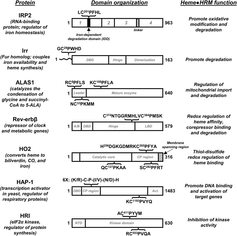
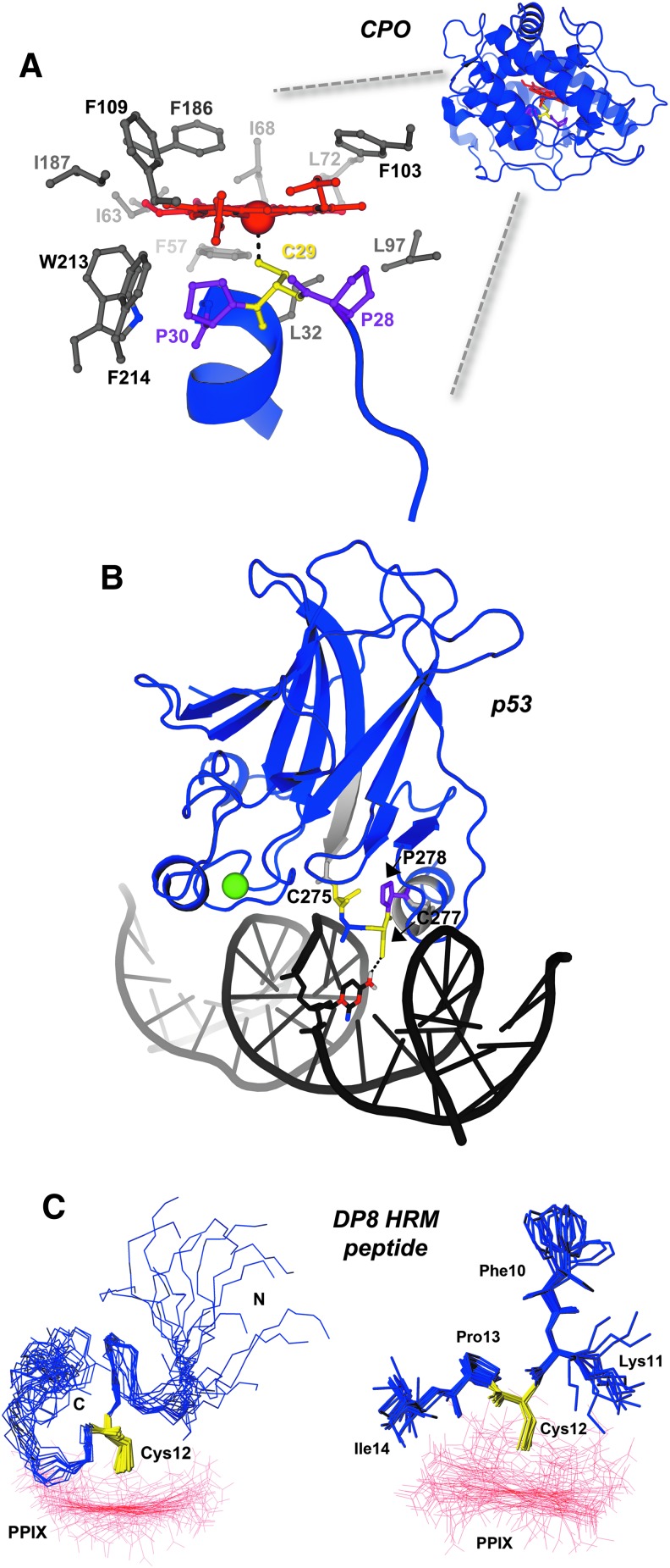
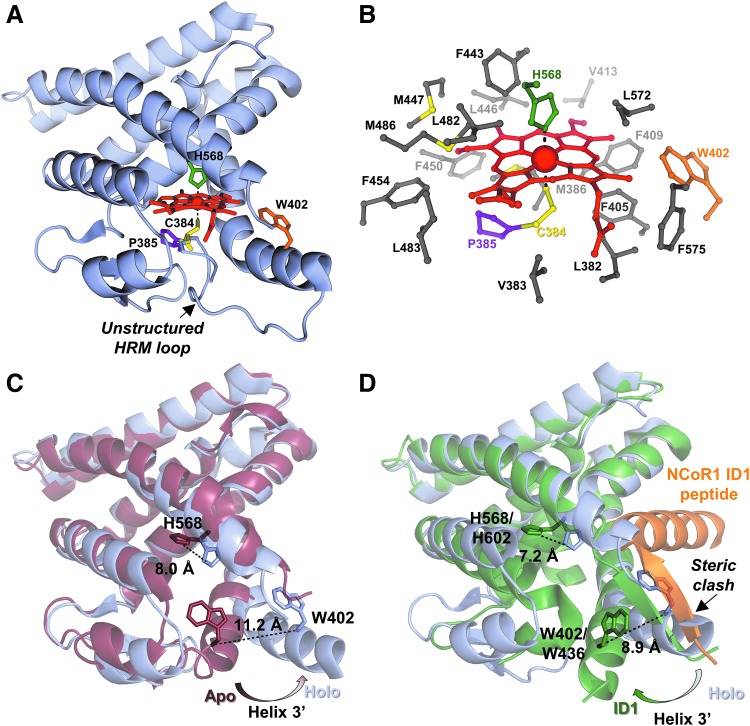
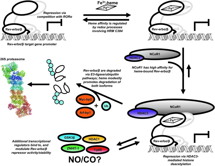
Significance: Heme binds to and serves as a cofactor for a myriad of proteins that are involved in diverse biological processes. Hemoproteins also exhibit varying modes of heme binding, suggesting that the protein environment contributes to the functional versatility of this prosthetic group. The subject of this review is a subset of hemoproteins that contain at least one heme regulatory motif (HRM), which is a short sequence containing a Cys-Pro core that, in many cases, binds heme with the Cys acting as an axial ligand. Recent Advances: As more details about HRM-containing proteins are uncovered, some underlying commonalities are emerging, including a role in regulating protein stability. Further, the cysteines of some HRMs have been shown to form disulfide bonds. Because the cysteines must be in the reduced, dithiol form to act as a heme axial ligand, heme binds at these sites in a redox-regulated manner, as demonstrated for heme oxygenase-2 (HO2) and Rev-erbβ.

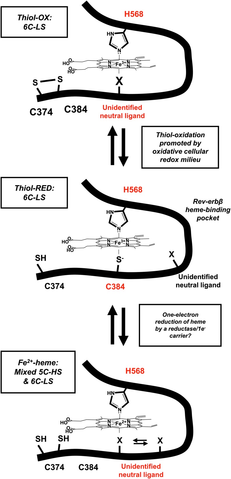
Critical issues: HRM-containing proteins have wide variations in heme affinity, utilize different axial ligand schemes, and exhibit differences in the ability to act as a redox sensor-all while having a wide variety of biological functions. Here, we highlight HO2 and Rev-erbβ to illustrate the similarities and differences between two hemoproteins that contain HRMs acting as redox sensors.

Future directions: HRMs acting as redox sensors may be applicable to other HRM-containing proteins as many contain multiple HRMs and/or other cysteine residues, which may become more evident as the functional significance of HRMs is probed in additional proteins.

Keywords: disulfide; heme; heme oxygenase; heme regulatory motif; nuclear receptor; redox regulation.


Figures

<b>FIG. 1.</b>
FIG. 1.
HRMs regulate the activity of enzymes, transcription factors, and an RNA regulatory protein. The modular structure of HRM-containing proteins is shown, along with the context and sequence of the HRMs. Domain organization (1–4) of IRP2 is based on its homology to aconitase (41, 74). Similarly, Irr is homologous to the Ferric Uptake Regulator (Fur) from Pseudomonas aeruginosa, and its domains are tentatively assigned as such; the N-terminal region of Irr that is distinct from Fur and contains the HRM is shown as a wavy line (29). The sequence of Rev-erbβ containing the HRM and redox-active Cys374 is shown; the A/B domain is hypervariable among members of the NR superfamily. For HO2, the sequence encompassing the heme ligand, His256 is shown in context to the nearby HRM. Hap-1 domain organization is based on functional studies (67); Act. refers to the domain that imparts transcription activation activity. NTD, or N-terminal domain of HRI is important for regulating quaternary structure (62). Delineation of domain boundaries are approximations. ALAS1, aminolevulinic acid synthase-1 nonspecific; Hap-1, heme activator protein-1; HO2, heme oxygenase isoform 2; HRM, heme regulatory motif; IRP2, iron regulatory protein 2; Irr, bacterial iron response regulator.
<b>FIG. 2.</b>
FIG. 2.
HRMs provide unique structural determinants to proteins and enzymes. (A) The overall structure of chloroperoxidase (PDB: 2ciw) shows that the HRM and Fe3+-heme cofactor are buried within the enzyme. A close-up view of the active site highlights the unique Pro-Cys-Pro HRM of CPO (prolines are shown as purple sticks, cysteine as yellow sticks), which positions the axial thiolate for heme ligation. Nonpolar residues within 4 Å of heme are shown as gray sticks. (B) p53, a transcription factor and tumor suppressor, is shown to be complexed with its cognate DNA promoter element (PDB: 1tsr). Residues flanking the HRM are shown as light gray cartoons to highlight the region, and a structural zinc atom is shown as a green sphere. (C) The NMR structure of an HRM-containing peptide from DP8 in complex with Ga3+-PPIX. Left panel, the 15 structures of lowest target function are shown with the peptide backbone as blue sticks, the HRM Cys12 in yellow sticks, and Ga3+-PPIX as red lines; the right panel depicts the inflexibility of the Cys-Pro core versus the side chains of surrounding residues. The structure serves as an excellent model demonstrating how the HRM Pro residue positions C-terminal residues away from heme, while poising the Cys-thiolate to act as a heme axial ligand. The structure of an IL-36α-derived HRM peptide in complex with Ga3+-PPIX recapitulates many of the features observed with the DP8 peptide (6). Figures in (C) are reprinted (adapted) with permission from Kühl et al. (50). Copyright (2013) American Chemical Society. CPO, chloroperoxidase; DP8, dipeptidyl peptidase 8; IL-36α, interleukin-36α; NMR, nuclear magnetic resonance; PPIX, protoporphyrin IX.
<b>FIG. 3.</b>
FIG. 3.
Structural insight into HRM-mediated redox regulation of heme binding to nuclear receptor, Rev-erbβ. (A) Structure of the Rev-erbβ LBD in complex with Fe3+-heme (holo-LBD, PDB: 3cqv). Heme binds in a 6-coordinate, low-spin complex with His (H568, green sticks) and Cys (C384, yellow sticks) axial ligands. C384 and P385 (purple sticks) comprise an HRM core that resides on a flexible, unstructured loop. W402 (orange sticks) provides hydrophobic contacts to heme, and its fluorescence is quenched on heme binding (unpublished observations). (B) The Rev-erbβ heme pocket is highly hydrophobic with nonpolar residues within 4 Å of heme shown as gray sticks (66). (C) Heme binding induces conformational changes of the LBD. Overlay of apo-LBD (dark red; PDB: 2v0v) and holo-LBD (light blue) structures aligned in Pymol; heme and the flexible HRM-harboring loop from the holo-LBD structure are omitted for clarity. The H3' helix swings out to accommodate the porphyrin ring, leading to a 11.2 Å shift of the W402 α-carbon. Further, conformational changes in helix 11 cause the τ-nitrogen of H568 to shift 8.0 Å, and assume its role as a Fe3+-heme ligand. (D) Pymol structural alignment of holo-LBD (light blue) versus apo-Rev-erbα LBD (dark green) in complex with an NCoR1 ID1 peptide (orange), PDB: 3n00; again, heme and the HRM loop are omitted for clarity. An anti-parallel beta sheet formed between an unraveled portion of Rev-erbα helix 11 and the NCoR1 peptide exhibits steric clash with helix H3' in the holo-LBD structure. Further, the heme axial ligand, H602 in Rev-erbα pivots away from the heme-binding pocket, suggesting that heme and NCoR1 would compete for different Rev-erb conformers. ID, interaction domain; LBD, ligand-binding domain; NCoR1, nuclear receptor compressor 1.
<b>FIG. 4.</b>
FIG. 4.
Heme-dependent repression and degradation of Rev-erbα/β. One mode of Rev-erbα/β repression is through competition with RORα for binding to its response element (ROR-RE) within the promoters of target genes. Rev-erbs can bind as monomers and homodimers to Rev-REs, although recent studies suggest that they may also form heterodimers (8, 13, 31). Although Fe3+-heme binds to Rev-erbβ with high affinity (Kd ≤0.1 nM), redox processes that cause dissociation of the Fe3+-heme axial thiolate ligand (including thiol-disulfide interconversion and Fe3+- to Fe2+-heme reduction) cause Kd to increase to ≥14 nM (11). Since the concentration of RH in the nucleus is ≤2.5 nM (30), loss of the HRM heme axial thiolate would cause dissociation of heme. Coimmunoprecipitation studies show that the NCoR1-HDAC3 complex has a high affinity for heme-bound Rev-erbα/β (70, 102); once bound, the complex leads to target gene repression via histone deacetylation. Additional regulators of Rev-erbβ repressor activity have been identified, including Tip60, an acetyl transferase, HDAC1, a histone deacetylase, and ZNHIT-1, a zinc-finger containing protein; GSK3β has also been shown to regulate the stability of the α-isoform (see main text for references); whether heme plays a role in regulating interactions of these proteins with Rev-erbs is unknown. Gaseous signaling molecules, NO and CO may also regulate the repressor function of Rev-erbs by binding to heme; however, additional studies are required to support this hypothesis. Although heme facilitates interaction of NCoR1 with Rev-erbs, it also appears to promote proteasomal degradation of both isoforms through a ubiquitin-dependent pathway involving E3-ligases Arf-bp1 and Myc-bp2. CO, carbon monoxide; HDAC1, histone deacetylase 1; NO, nitric oxide; RH, regulatory heme; RORα, RAR-related orphan receptor α.
<b>FIG. 5.</b>
FIG. 5.
Rev-erbβ undergoes redox-mediated heme ligand switching. The thiol-reduced form of the Rev-erbβ LBD (thiol-RED; C374 and C384 exist as thiols/thiolates) binds Fe3+-heme in a 6C-LS complex with H568 and C384 axial ligands and a Kd of ∼100 pM. Under oxidizing conditions, C384 forms a disulfide with neighboring C374 (thiol-OX), leading to a 6C-LS system where an unidentified neutral ligand assumes the position once occupied by the thiolate. Thiol-OX has a Kd for Fe3+-heme of ≥14 nM, well above the nuclear RH level of ≤2.5 nM, suggesting that thiol-OX would exist as an apoprotein in the nucleus. Our working hypothesis is that C374–C384 thiol-disulfide interconversion is in equilibrium with the GSH:GSSG or cysteine:cystine couples and reflects the redox poise of the nucleus. Similarly, the one-electron reduction of Fe3+- to Fe2+-heme is accompanied by dissociation of the HRM heme axial thiolate, leading to a mixed 5C-HS/6C-LS system where an unidentified neutral ligand (presumably the same ligand involved in the thiol-RED/OX ligand switch) is loosely associated with heme. Although it is unclear what heme redox state is relevant to Rev-erbβ in the cell, it is intriguing to consider one-electron reductants that could interface with Rev-erbβ, such as ferredoxins or cytochrome P450 reductase, although nuclear localization of the reductant is presumably required. 5C-HS, five-coordinate high spin; 6C-LS, six-coordinate low-spin.
<b>FIG. 6.</b>
FIG. 6.
Spectral characterization of HO2 in the Fe3+-heme-bound forms. (A) Absorbance spectra of 5 μM Fe3+-heme-bound forms of HO2core (), HO2tailR (formula image
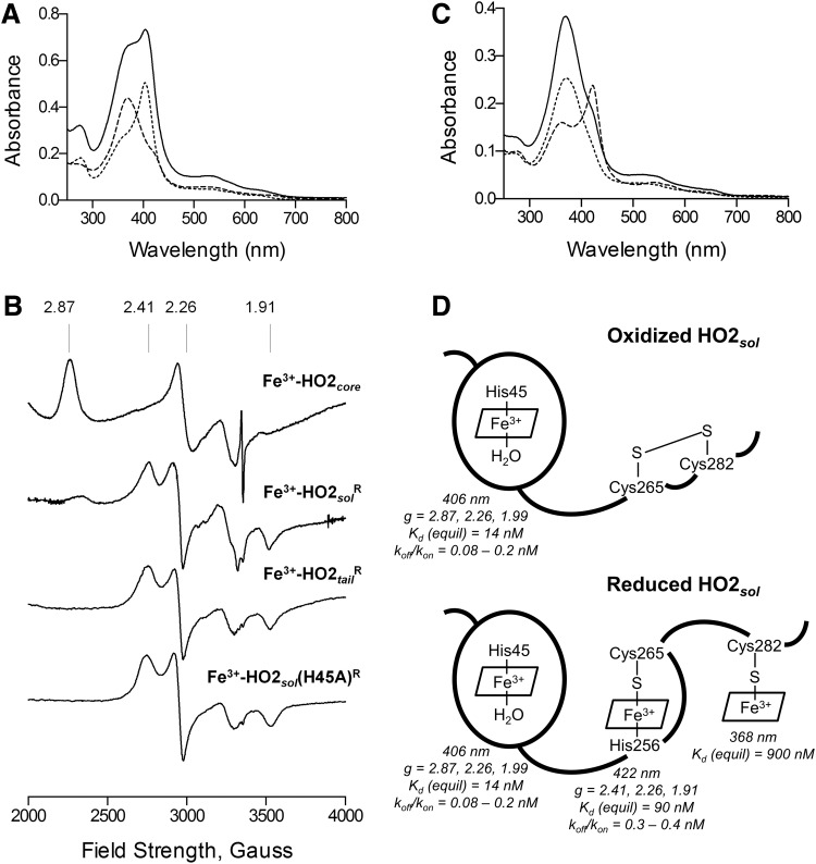
), and HO2solR (—) in 50 mM Tris (pH 8.0) and 50 mM KCl at 20°C. (B) EPR spectra of the Fe3+-heme-bound forms of HO2core, HO2solR, HO2tailR, and the His45Ala variant of HO2solR. The g values are indicated above the spectra. (C) Absorbance spectra of 5 μM Fe3+-heme-bound forms of HO2tailR (—), HO2tail(C282A)R (formula image), and HO2tail(C265A)R () in 50 mM Tris (pH 8.0) and 50 mM KCl at 20°C. (D) A model of heme binding to the oxidized form of HO2sol, which has a single heme-binding site due to the participation of Cys265 and Cys282 in a disulfide bond, and the reduced form of HO2sol, which has three heme-binding sites. Below each heme-binding site are the characteristics of that site, including the maximum absorbance, the g-values, the Kd values obtained by equilibrium titrations [Kd (equil)], and the koff/kon values obtained by kinetic measurements. The figures in (A–C) are reprinted (adapted) with permission from Fleischhacker et al. (20). Copyright (2015) American Chemical Society. EPR, electron paramagnetic resonance; HO2core, HO2 spanning residues 1-248; HO2R, HO2 in the disulfide bond-reduced form; HO2sol, HO2 spanning residues 1-288; HO2tail, HO2 spanning residues 213-288. EPR, electron paramagnetic resonance; HO2core, HO2 spanning residues 1-248; HO2R, HO2 in the disulfide bond-reduced form; HO2sol, HO2 spanning residues 1-288; HO2tail, HO2 spanning residues 213-288.

References

    1. Akashi M. and Takumi T. The orphan nuclear receptor RORα regulates circadian transcription of the mammalian core-clock Bmal1. Nat Struct Mol Biol 12: 441–448, 2005 - PubMed
    1. Bagai I, Sarangi R, Fleischhacker AS, Sharma A, Hoffman BM, Zuiderweg ERP, and Ragsdale SW. Spectroscopic studies reveal that the heme regulatory motifs of heme oxygenase-2 are dynamically disordered and exhibit redox-dependent interaction with heme. Biochemistry 54: 2693–2708, 2015 - PMC - PubMed
    1. Barr I, Smith AT, Senturia R, Chen YQ, Scheidemantle BD, Burstyn JN, and Guo F. DiGeorge critical region 8 (DGCR8) is a double-cysteine-ligated heme protein. J Biol Chem 286: 16716–16725, 2011 - PMC - PubMed
    1. Bianchetti CM, Yi L, Ragsdale SW, and Phillips GN., Jr Comparison of apo- and heme-bound crystal structures of a truncated human heme oxygenase-2. J Biol Chem 282: 37624–37631, 2007 - PMC - PubMed
    1. Bohle DS, Dodd EL, Pinter TBJ, and Stillman MJ. Soluble diamagnetic model for malaria pigment: coordination chemistry of gallium(III)protoporphyrin-IX. Inorg Chem 51: 10747–10761, 2012 - PubMed

Publication types

MeSH terms

LinkOut - more resources