Review
. 2018 Feb;18(2):91-104.
doi: 10.1038/nri.2017.112. Epub 2017 Oct 9.

Immune checkpoint blockade in infectious diseases

Affiliations
Review

Immune checkpoint blockade in infectious diseases

Michelle N Wykes et al. Nat Rev Immunol. 2018 Feb.

Abstract

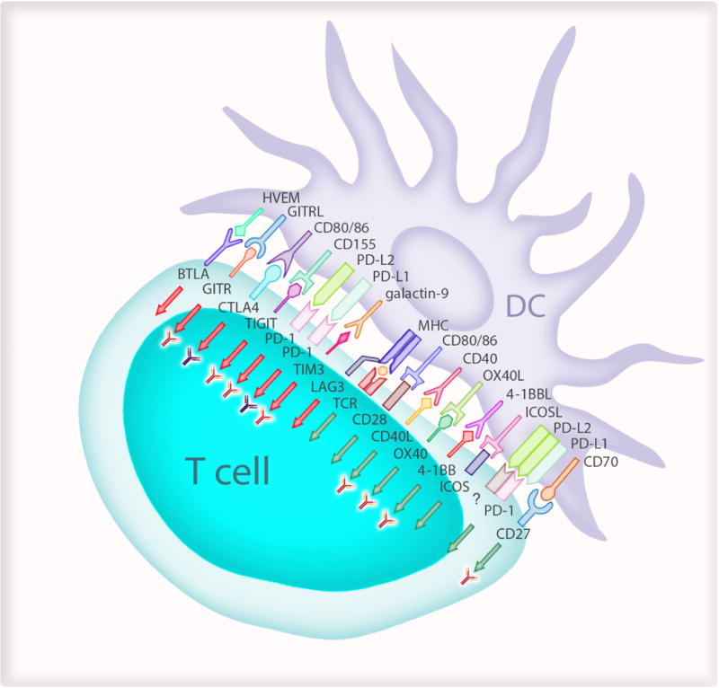
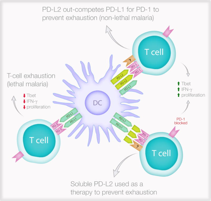
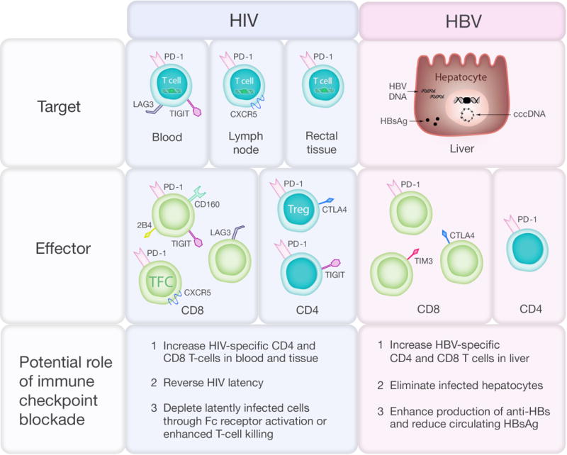
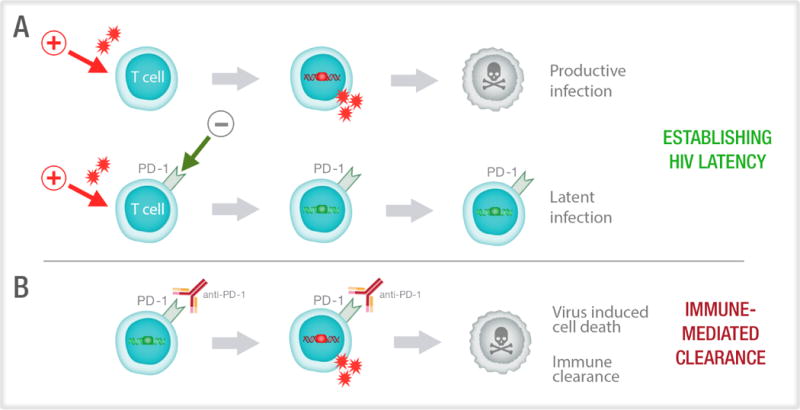
The upregulation of immune checkpoint molecules, such as programmed cell death protein 1 (PD1) and cytotoxic T lymphocyte antigen 4 (CTLA4), on immune cells occurs during acute infections, such as malaria, as well as during chronic persistent viral infections, including HIV and hepatitis B virus. These pathways are important for preventing immune-driven pathology but can also limit immune-mediated clearance of the infection. The recent success of immune checkpoint blockade in cancer therapy suggests that targeting these pathways would also be effective for preventing and treating a range of infectious diseases. Here, we review our current understanding of immune checkpoint pathways in the pathogenesis of infectious diseases and discuss the potential for therapeutically targeting these pathways in this setting.


Figures

Figure 1
Figure 1. Interactions that regulate T cell responses
Antigen presenting cells such as dendritic cells (DCs) regulate T cell response to specific pathogens or antigens from malignant cells. The T cell receptors (TCR) on antigen-specific T cells first recognise their cognate antigen via the major histocompatibility complex (MHC) molecules on antigen presenting cells. This step has to be followed by signals to CD28 on T cells from CD80 on the APC and is described as “signal 2”. Several different ligands on DCs then provide signals to T cells which decide the quality and duration of the effector response (green arrows). These include CD40/CD40 ligand (CD40L); OX40/OX40 ligand (OX40L); 4-1BB (CD137)/4-1BB ligand (41BBL; CD137 Ligand); ICOS (Inducible T-cell COStimulator; CD278)/ICOS Ligand (ICOS-L); CD27/CD70. There are also signals to suppress immune responses (red arrows) to maintain self tolerance and limit the duration of immune responses to minimize bystander damage to host tissue. These include LAG3 (lymphocyte activation gene 3); MHC class II; TIM3 (T cell immunoglobulin and mucin-domain containing-3; HAVCR2 in humans)/galectin-9; PD-1 (programmed cell death-1)/PD-L1 (programmed cell death-1-ligand 1) and PD-L2 (programmed cell death-1-ligand 2); TIGIT (T cell immunoreceptor with Ig and ITIM domains)/CD155; CTLA4 (cytotoxic T-lymphocyte-associated protein 4)/CD86 or CD80; GITR (Glucocorticoid-induced TNFR-related protein)/GITR-L (GITR-ligand) and BTLA (B and T lymphocyte attenuator)/HVEM (Herpesvirus entry mediator). Antibody symbol represents pathways being tested in current clinical trials. The “?” refers to an unknown receptor which “activates” T cells. The “red” antibodies indicate pathways undergoing clinical trials for cancer and the “dark coloured” antibodies indicate clinical use.
Figure 2
Figure 2. PD-L2 protects against lethal malaria and has translational potential
The expression of PD-L2 on dendritic cells determines effector T cell function following a PD-1/PD-L1 interaction. During non-lethal malaria (top right), DCs express PD-L2 which inhibits the immunosuppressive PD-1/PD-L1 interaction while interacting with an unknown receptor (?) to improve T cell functions. This leads to protective immunity characterised by increased T-box transcription factor TBX21 (Tbet) expression, increase interferon-γ (IFN-γ) secretion and better proliferation in response to the parasite. In contrast, during lethal malaria (left), PD-L2 expression is low or absent and this allows the immunosuppressive PD-1/PD-L1 interaction to generate exhausted T cells which do not express Tbet, do not secrete IFN-γ and cannot proliferate in response to the parasite. Soluble PD-L2 administered to mice infected with lethal malaria (lower right) can prevent T cell exhaustion.
Figure 3
Figure 3. Immune checkpoint protein expression in HIV and HBV infection and potential effects of immune checkpoint blockade
In HIV infection, the virus persists on antiretroviral therapy (ART) in latently infected CD4+ T-cells that contain integrated provirus (green box) and express PD-1 and other immune checkpoint markers in blood, lymph node and rectal tissue. Expression of immune checkpoint markers on total and HIV-specific CD4+ and CD8+ T cell subsets include central memory (CM), effector memory (EM), T follicular cytotoxic (TFC) and T regulatory (Treg) T cells is associated with T-cell exhaustion and reduced T-cell function. In HBV infection, HBV persists on treatment as extrachromosomal closed covalent circular (ccc) DNA and integrated HBV DNA (black box) and there is ongoing production of HBV surface antigen (HBsAg). Increased expression of immune checkpoint markers on CD8+ T cells and increased expression of PD-1 on CD4+ T-cells reduce T cell function.
Figure 4
Figure 4. Proposed role of PD-1 in the establishment and reversal of HIV latency
(A) HIV preferentially infects activated CD4+ T cells which have been stimulated via T cell receptor engagement or a mitogen (red arrow). Following HIV integration, the productively infected cell (red box) usually dies by virus mediated cytolysis. Up-regulation of immune checkpoint markers such as PD-1, could potentially limit T-cell activation favouring latent over productive infection, where there is integration (green box) but no virus production. (B) Latently infected cells express immune checkpoint markers, including PD-1. The administration of anti-PD-1, or other immune checkpoint blockers, leads to activation of the T cells and increased expression of transcription factors that can enhance production of virus from latency. This leads to either immune mediated clearance or virus induced cell death.

Similar articles

Cited by

References

    1. Baumeister SH, Freeman GJ, Dranoff G, Sharpe AH. Coinhibitory Pathways in Immunotherapy for Cancer. Annu Rev Immunol. 2016;34:539–573. doi: 10.1146/annurev-immunol-032414-112049. A comprehensive review on inhibitory pathways. - DOI - PubMed
    1. Pardoll DM. The blockade of immune checkpoints in cancer immunotherapy. Nat Rev Cancer. 2012;12:252–264. doi: 10.1038/nrc3239. An excellent review on checkpoint inhibition in the context of cancer. - DOI - PMC - PubMed
    1. Wherry EJ. T cell exhaustion. Nat Immunol. 2011;12:492–499. A definitive review of T cell exhaustion. - PubMed
    1. Larkin J, et al. Combined Nivolumab and Ipilimumab or Monotherapy in Untreated Melanoma. N Engl J Med. 2015;373:23–34. doi: 10.1056/NEJMoa1504030. - DOI - PMC - PubMed
    1. Brahmer JR, et al. Phase I study of single-agent anti-programmed death-1 (MDX-1106) in refractory solid tumors: safety, clinical activity, pharmacodynamics, and immunologic correlates. J Clin Oncol. 2010;28:3167–3175. doi: 10.1200/JCO.2009.26.7609. - DOI - PMC - PubMed

Publication types

MeSH terms

Substances