. 2018 Jun;19(6):1414-1426.
doi: 10.1111/mpp.12624. Epub 2018 Feb 9.

Disruption of calcineurin catalytic subunit (cnaA) in Epichloë festucae induces symbiotic defects and intrahyphal hyphae formation

Affiliations

Disruption of calcineurin catalytic subunit (cnaA) in Epichloë festucae induces symbiotic defects and intrahyphal hyphae formation

Milena Mitic et al. Mol Plant Pathol. 2018 Jun.

Abstract

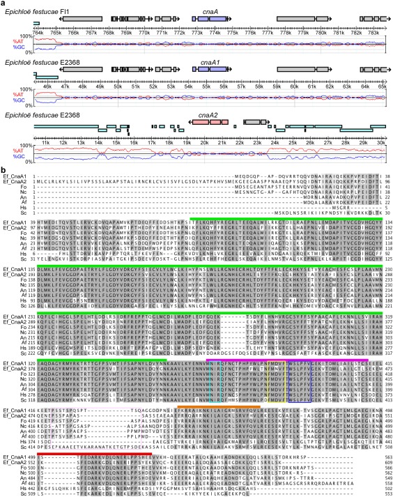
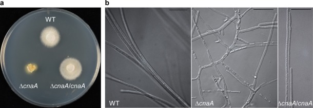
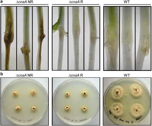
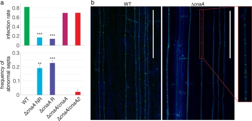
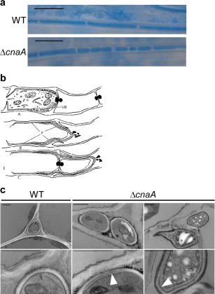
Calcineurin is a conserved calcium/calmodulin-dependent protein phosphatase, consisting of a catalytic subunit A and a regulatory subunit B, which is involved in calcium-dependent signalling and regulation of various important cellular processes. In this study, we functionally characterized the catalytic subunit A (CnaA) of the endophytic fungus Epichloë festucae which forms a symbiotic association with the grass host Lolium perenne. We deleted the CnaA-encoding gene cnaA in E. festucae and examined its role in hyphal growth, cell wall integrity and symbiosis. This ΔcnaA strain had a severe growth defect with loss of radial growth and hyper-branched hyphae. Transmission electron microscopy and confocal microscopy analysis of the mutant revealed cell wall defects, aberrant septation and the formation of intrahyphal hyphae, both in culture and in planta. The mutant strain also showed a reduced infection rate in planta. The fluorescence of mutant hyphae stained with WGA-AF488 was reduced, indicating reduced chitin accessibility. Together, these results show that E. festucae CnaA is required for fungal growth, maintaining cell wall integrity and host colonization.

Keywords: CnaA; Epichloë; calcineurin; hyphal growth.


Figures

Figure 1
Figure 1
Multiple copies of cnaA in Epichloë festucae. (a) Physical maps of the cnaA loci in E. festucae Fl1 and E2368. The percentage AT/GC content (red/blue traces) and repetitive elements (cyan bars) are shown. Scale bar indicates the position within host contigs. (b) Alignment of the deduced amino acid sequences of cnaA1 and cnaA2 from E. festucae and cnaA from Fusarium oxysporum (Fo, XP018243626), Neurospora crassa (Nc, XP011394598), Aspergillus nidulans (An, P48457), Aspergillus fumigatus (Af, XP753703), Saccharomyces cerevisiae (Sc, NP013537) and human (Hs, AAB23769). Conserved domains are shown with coloured bars: catalytic (green), calcineurin B‐binding (purple), calmodulin‐binding (orange) and autoinhibitory region (red).
Figure 2
Figure 2
Deletion of Epichloë festucae Fl1 cnaA causes severe growth defects in culture. (a) Colony morphology of E. festucae Fl1 wild‐type (WT), cnaA deletion strain (ΔcnaA) and ΔcnaA strain complemented with WT copy of E. festucae Fl1 cnaAcnaA/cnaA) grown on potato dextrose agar for 6 days. (b) Comparison of differential interference contrast image of ΔcnaA culture showing highly deregulated growth pattern with increased branching and altered hyphal morphology compared with the wild‐type (WT) and complementation strain (ΔcnaA/cnaA). Bar, 20 μm.
Figure 3
Figure 3
Deletion of Epichloë festucae Fl1 cnaA causes defects in septation and the distribution of cell wall components. Confocal images of E. festucae Fl1 wild‐type (WT), ΔcnaA and ΔcnaA/cnaA cultures stained with calcofluor white, which binds to chitin, showing a loss of the WT staining pattern in the ΔcnaA strain. Bar, 20 μm.
Figure 4
Figure 4
Deletion of Epichloë festucae Fl1 cnaA causes the formation of intrahyphal hyphae. (a) Transmission electron micrographs of E. festucae Fl1 wild‐type (WT) and ΔcnaA strains showing extensive formation of intrahyphal cell walls in the mutant hyphae (indicated by yellow arrowheads) and abnormal septa (indicated by white arrowheads). Red arrowheads indicate disrupted outer cell wall. Bar, 2 μm. (b) Reproduction of a drawing from Calonge (1968) showing intrahyphal hyphae breaking out from the mother hypha of Sclerotinia fructigena. This schematic diagram captures the transmission electron micrographs of the hyphal structures observed in the ΔcnaA strain.
Figure 5
Figure 5
The duplicate cnaA genes of Epichloë festucae E2368 are functionally redundant. (a) Colony morphology of E. festucae E2368 wild‐type (WT), ΔcnaA1#17 (ΔcnaA1) and ΔcnaA2#27 (ΔcnaA2) cultures grown on potato dextrose agar for 6 days. (b) Colony morphology of E. festucae Fl1 ΔcnaA complemented with the WT copy of E. festucae E2368 cnaA2cnaA/cnaA2) grown on potato dextrose agar for 6 days.
Figure 6
Figure 6
Deletion of Epichloë festucae Fl1 cnaA elicits a host defence response in Lolium perenne seedlings. (a) Light micrographs of L. perenne seedlings at 10 days post‐inoculation with non‐remediated (ΔcnaA NR) and remediated (ΔcnaA R) cultures of Fl1 ΔcnaA and wild‐type (WT). (b) Epichloë festucae grow outs from L. perenne plants infected with non‐remediated ΔcnaAcnaA NR), remediated ΔcnaAcnaA R) and wild‐type (WT) Fl1.
Figure 7
Figure 7
Epichloë festucae Fl1 ΔcnaA shows a reduced infection rate and abnormal septal morphology and patterning in planta. (a) Deletion of cnaA in E. festucae reduces the plant infection rate (upper panel). Deletion of cnaA in E. festucae increases the number of abnormal septa (lower panel). (b) Confocal micrographs of aniline blue‐ and wheat germ agglutinin‐conjugated Alexa Fluor®488 (WGA‐AF488)‐stained hyphae of wild‐type (WT) and ΔcnaA mutant in planta showing irregular septa formation. Bar, 100 μm.
Figure 8
Figure 8
Deletion of cnaA in Epichloë festucae Fl1 causes the formation of intrahyphal hyphae in planta. (a) Light micrographs of aniline blue‐stained hyphae of ΔcnaA and wild‐type (WT) showing constricted hyphae and increased septa formation in ΔcnaA relative to WT. Bar, 10 μm. (b) Reproduction of a drawing from Calonge (1968) showing intrahyphal hyphae breaking out from the mother hypha of Sclerotinia fructigena. This schematic diagram captures the light micrograph image of the hyphal structures observed in planta for E. festucae ΔcnaA hyphae. (c) Transmission electron micrographs of wild‐type (WT) and ΔcnaA mutant in planta showing duplication of the cell wall in mutant hyphae, suggesting the presence of intrahyphal hyphae. White arrowheads point to a double cell wall. Bar, 1 μm.

Similar articles

Cited by

References

    1. Alshahni, M.M. , Shimizu, K. , Yoshimoto, M. , Yamada, T. , Nishiyama, Y. , Arai, T. and Makimura, K. (2016) Genetic and phenotypic analyses of calcineurin A subunit in Arthroderma vanbreuseghemii . Med. Mycol. 54, 207–218. - PubMed
    1. Becker, M. , Becker, Y. , Green, K. and Scott, B. (2016) The endophytic symbiont Epichloë festucae establishes an epiphyllous net on the surface of Lolium perenne leaves by development of an expressorium, an appressorium‐like leaf exit structure. New Phytol. 211, 240–254. - PMC - PubMed
    1. Becker, Y. , Eaton, C.J. , Brasell, E. , May, K.J. , Becker, M. , Hassing, B. , Cartwright, G.M. , Reinhold, L. and Scott, B. (2015) The fungal cell‐wall integrity MAPK cascade is crucial for hyphal network formation and maintenance of restrictive growth of Epichloë festucae in symbiosis with Lolium perenne . Mol. Plant–Microbe Interact. 28, 69–85. - PubMed
    1. Blankenship, J.R. , Wormley, F.L. , Boyce, M.K. , Schell, W.A. , Filler, S.G. , Perfect, J.R. and Heitman, J. (2003) Calcineurin is essential for Candida albicans survival in serum and virulence. Eukaryot. Cell, 2, 422–430. - PMC - PubMed
    1. Bowman, S.M. , Piwowar, A. , Al Dabbous, M. , Vierula, J. and Free, S.J. (2006) Mutational analysis of the glycosylphosphatidylinositol (GPI) anchor pathway demonstrates that GPI‐anchored proteins are required for cell wall biogenesis and normal hyphal growth in Neurospora crassa . Eukaryot. Cell, 5, 587–600. - PMC - PubMed

LinkOut - more resources