. 2017 Nov 1;139(43):15377-15384.
doi: 10.1021/jacs.7b07147. Epub 2017 Oct 23.

Self-Assembling Ability Determines the Activity of Enzyme-Instructed Self-Assembly for Inhibiting Cancer Cells

Affiliations

Self-Assembling Ability Determines the Activity of Enzyme-Instructed Self-Assembly for Inhibiting Cancer Cells

Zhaoqianqi Feng et al. J Am Chem Soc. .

Abstract

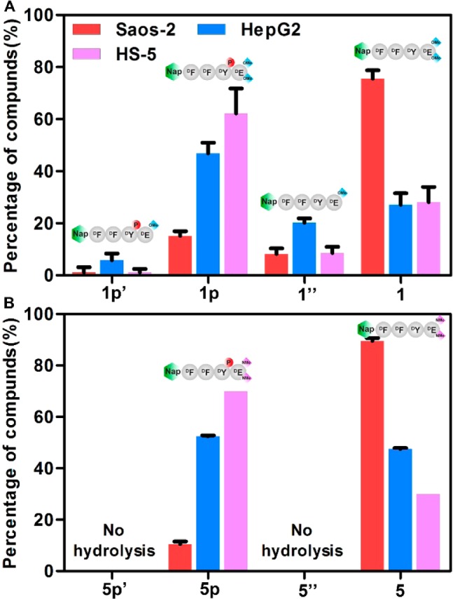
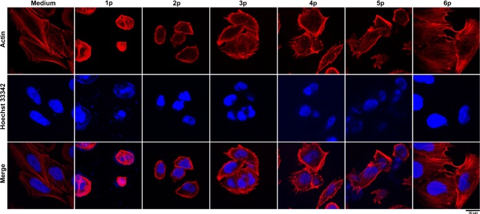
Enzyme-instructed self-assembly (EISA) represents a dynamic continuum of supramolecular nanostructures that selectively inhibits cancer cells via simultaneously targeting multiple hallmark capabilities of cancer, but how to design the small molecules for EISA from the vast molecular space remains an unanswered question. Here we show that the self-assembling ability of small molecules controls the anticancer activity of EISA. Examining the EISA precursor analogues consisting of an N-capped d-tetrapeptide, a phosphotyrosine residue, and a diester or a diamide group, we find that, regardless of the stereochemistry and the regiochemistry of their tetrapeptidic backbones, the anticancer activities of these precursors largely match their self-assembling abilities. Additional mechanistic studies confirm that the assemblies of the small peptide derivatives result in cell death, accompanying significant rearrangement of cytoskeletal proteins and plasma membranes. These results imply that the diester or diamide derivatives of the d-tetrapeptides self-assemble pericellularly, as well as intracellularly, to result in cell death. As the first case to correlate thermodynamic properties (e.g., self-assembling ability) of small molecules with the efficacy of a molecule process against cancer cells, this work provides an important insight for developing a molecular dynamic continuum for potential cancer therapy, as well as understanding the cytotoxicity of pathogenic assemblies.


Conflict of interest statement

The authors declare no competing financial interest.

Figures

Scheme 1
Scheme 1. Molecular Structures of the Precursors and the Correlation between the Ability for Self-Assembly of Small Molecules and Anticancer Activity
Figure 1
Figure 1
TEM images of the nanostructures formed by 0.5 wt % 1p6p in pH 7.4 water, before and after adding ALP (2 U/mL) (scale bar, 100 nm).
Figure 2
Figure 2
Cmc values of precursors 1p6p and their corresponding dephosphorylated peptide derivatives 16.
Figure 3
Figure 3
Correlation between the self-assembling ability (−ΔG0) and anticancer activity (pIC50) of EISA molecules against Saos-2 cells.
Figure 4
Figure 4
(A) Intensity of static light scattering (SLS) of the solutions of 1p, 5p, and 7p (5–50 μM) before and after adding ALP (1 U/mL) for 12 h in pH 7.4 PBS buffer (light-scattering angle = 60°). (B) The cell viability of Saos-2 cells treated with 1p, 5p, or 7p (5–50 μM) for 24 h.
Figure 5
Figure 5
IC50 (72 h) of 1p or 5p against Saos-2 cells, MCF-7 cells, T98G cells, or HS-5 cells.
Figure 6
Figure 6
(A) Percentage of compounds 1p′ (hydrolysis product of 1p), 1p, 1″, and 1 after incubating 1p (200 μM) with Saos-2 cells, HepG2 cells, or HS-5 cells. (B) Percentage of compounds 5p′ (hydrolysis product of 5p), 5p, 5″, and 5 after incubating 5p (200 μM) with Saos-2 cells, HepG2 cells, or HS-5 cells. The ratios were determined using HPLC and LC–MS. Cells were treated for 24 h.
Figure 7
Figure 7
CLSM images of Saos-2 cells stained with Alexa Fluor 633 Phalloidin (F-actin, red) and Hoechst (nuclei, blue) after the treatment of culture medium, 1p, 2p, 3p, 4p, 5p, or 6p for 12 h. Scale bars = 20 μm.
Figure 8
Figure 8
CLSM images of Saos-2 cells stained with tubulin tracker (green) and Hoechst (nuclei, blue) after the treatment of culture medium, 1p, 2p, 3p, 4p, 5p, and 6p for 12 h. Scale bars = 20 μm.

Similar articles

Cited by

References

    1. Hopkins A. L.; Groom C. R. Nat. Rev. Drug Discovery 2002, 1, 727.10.1038/nrd892. - DOI - PubMed
    1. Verdine G. L.; Walensky L. D. Clin. Cancer Res. 2007, 13, 7264.10.1158/1078-0432.CCR-07-2184. - DOI - PubMed
    1. Makley L. N.; Gestwicki J. E. Chem. Biol. Drug Des. 2013, 81, 22.10.1111/cbdd.12066. - DOI - PMC - PubMed
    1. Carlier M. F. Philos. Trans. R. Soc., B 1992, 336, 93.10.1098/rstb.1992.0048. - DOI - PubMed
    1. Li J.; Kuang Y.; Gao Y.; Du X.; Shi J.; Xu B. J. Am. Chem. Soc. 2013, 135, 542.10.1021/ja310019x. - DOI - PMC - PubMed
    2. Yang Z.; Liang G.; Guo Z.; Guo Z.; Xu B. Angew. Chem., Int. Ed. 2007, 46, 8216.10.1002/anie.200701697. - DOI - PubMed

Publication types