. 2017 Nov 1;127(11):4124-4135.
doi: 10.1172/JCI94495. Epub 2017 Oct 9.

Caspase-11-mediated endothelial pyroptosis underlies endotoxemia-induced lung injury

Affiliations

Caspase-11-mediated endothelial pyroptosis underlies endotoxemia-induced lung injury

Kwong Tai Cheng et al. J Clin Invest. .

Abstract

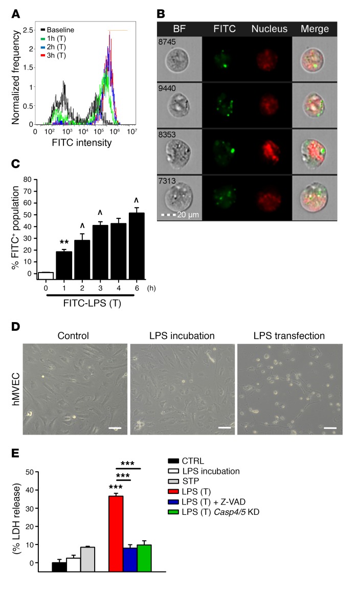
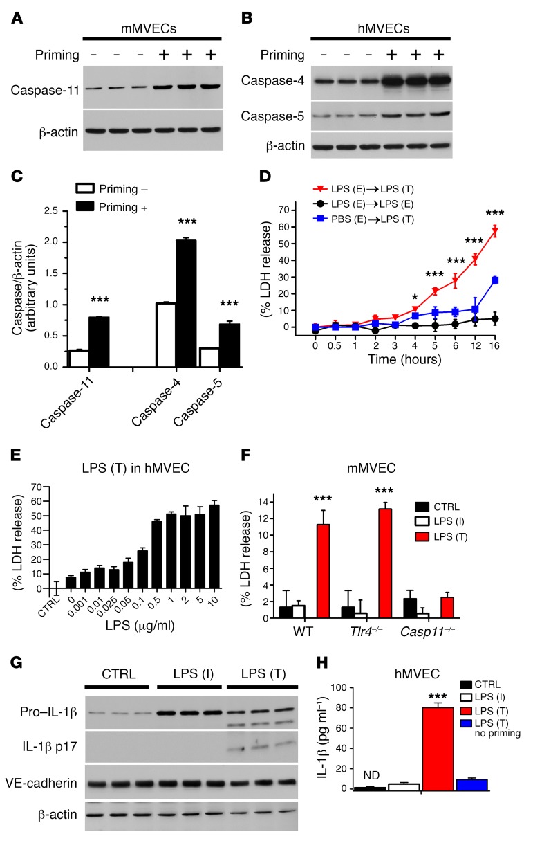
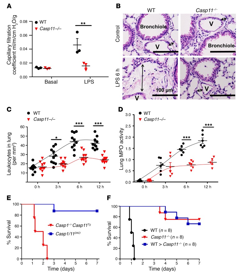
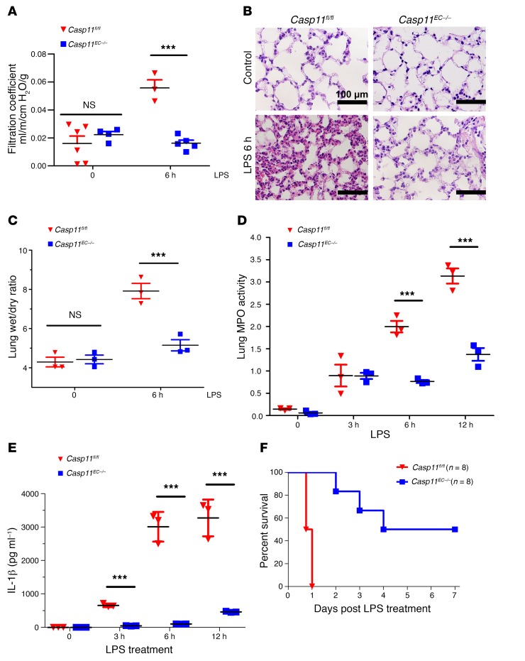
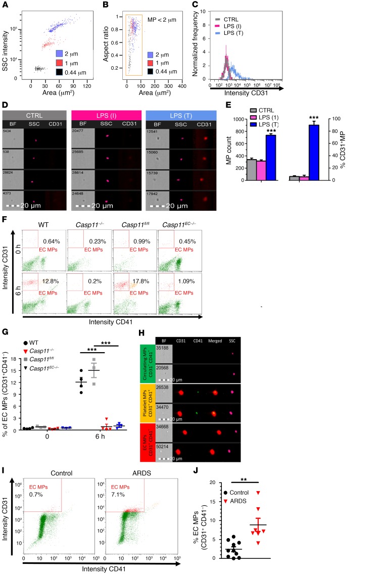
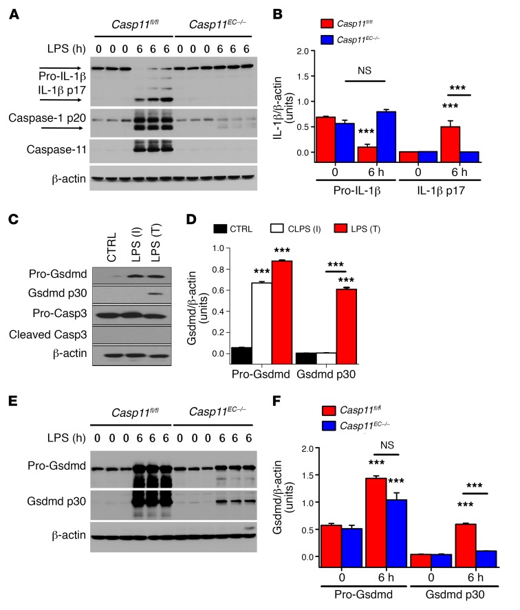
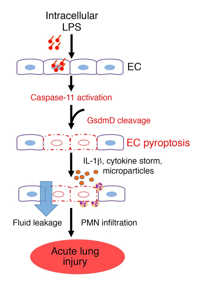
Acute lung injury is a leading cause of death in bacterial sepsis due to the wholesale destruction of the lung endothelial barrier, which results in protein-rich lung edema, influx of proinflammatory leukocytes, and intractable hypoxemia. Pyroptosis is a form of programmed lytic cell death that is triggered by inflammatory caspases, but little is known about its role in EC death and acute lung injury. Here, we show that systemic exposure to the bacterial endotoxin lipopolysaccharide (LPS) causes severe endothelial pyroptosis that is mediated by the inflammatory caspases, human caspases 4/5 in human ECs, or the murine homolog caspase-11 in mice in vivo. In caspase-11-deficient mice, BM transplantation with WT hematopoietic cells did not abrogate endotoxemia-induced acute lung injury, indicating a central role for nonhematopoietic caspase-11 in endotoxemia. Additionally, conditional deletion of caspase-11 in ECs reduced endotoxemia-induced lung edema, neutrophil accumulation, and death. These results establish the requisite role of endothelial pyroptosis in endotoxemic tissue injury and suggest that endothelial inflammatory caspases are an important therapeutic target for acute lung injury.


Conflict of interest statement

Conflict of interest: The authors have declared that no conflict of interest exists.

Figures

Figure 1
Figure 1. Intracellular LPS induces EC pyroptosis via activation of inflammatory caspases in mice and humans.
(A) Flow cytometry histograms and (B) representative cytometry images of hMVECs transfected with 2 μg/ml FITC-labeled LPS (O111:B4) for 3 hours. LPS fluorescence in green and nuclear staining in red show that LPS crossed the EC plasma membrane. Scale bar: 20 μm. BF, bright field. (C) Time course of hMVEC intracellular FITC-LPS fluorescence in the presence of a transfection (T) reagent. Results are shown as mean ± SEM. n = 5. **P < 0.01 from baseline; ^P < 0.01 from previous group using ANOVA. (D) Phase-contrast micrographs of control hMVECs after a 16-hour period of LPS (2 μg/ml) incubation and after a 16-hour period of LPS transfection (2 μg/ml) show that LPS transfection, but not LPS incubation, induced lytic cell death. Scale bars: 100 μm. (E) Release of LDH showed that LPS incubation (2 μg/ml) for 16 hours or staurosporine-induced (STP-induced) apoptosis resulted in minimal LDH release, whereas LPS transfection (2 μg/ml) led to marked cell lysis. Cells were primed with an initial exposure to LPS (500 ng/ml for 3 hours). Cell lysis was blocked by the pan-caspase inhibitor Z-VAD-FMK or knockdown (KD) of the human inflammatory caspases 4 and 5 (Casp4/5). Results are shown as mean ± SEM. n = 5. ***P < 0.001 using ANOVA.
Figure 2
Figure 2. TLR4-mediated LPS signaling is required for activation of endothelial pyroptosis.
Immunoblot analysis of the inflammatory caspase-11 in mMVECs (A) and the inflammatory caspases 4 and 5 in hMVECs (B) in the presence or absence of priming with extracellular 500 ng/ml LPS for 3 hours prior to transfecting the cells with 2 μg/ml LPS for 16 hours. (C) Quantification of the inflammatory caspase expression shows significant upregulation of caspases 4 and 5 in human ECs and caspase-11 in mouse ECs with priming. Statistics obtained from Student’s 2-tailed t test. (D) hMVECs were first primed with extracellular (E) LPS or PBS, then transfected (T) with LPS or only incubated (I) with extracellular LPS. (E) Dose dependence of EC lysis in hMVECs induced by intracellular LPS. (F) LDH release by mMVEC isolated from WT, Tlr4–/–, and Casp11–/– mice after internalization of LPS for 16 hours. CTRL, control. (G) Western blot and (H) ELISA detection of mature IL-1β in hMVEC lysates and culture supernatants 16 hours after endothelial transfection with LPS (2 μg/ml) or without LPS transfection. (H) There was a significant amount of mature IL-1β release when ECs were primed with extracellular LPS and subsequently exposed to intracellular LPS for 16 hours. Results are shown as mean ± SEM. n ≥ = 5. *P < 0.05; ***P < 0.001. All statistics except C obtained from ANOVA.
Figure 3
Figure 3. Requirement for caspase-11 expressed in nonhematopoietic cells in mediating endotoxemia-induced acute lung vascular injury and mortality.
(A) Lung microvessel filtration coefficient (a measure of lung vascular permeability) was determined in WT and Casp11–/– mice, and the data points depicting individual mice are shown. Mice were exposed to systemic LPS (40 mg/kg i.p.) for 6 hours. Statistics obtained from 2-tailed Student’s t test.(B) H&E-stained cross section of the lung from control mice and LPS-exposed mice at 6 hours shows interstitial edema (indicated by arrows) in WT but not in Casp11–/– mice. Scale bars: 100 μm. V, lung microvasculature. Images are representative of 5 animals. Quantitative analysis for leukocyte infiltration in lungs (C) and lung tissue MPO activity (D). *P < 0.05; **P < 0.01; ***P < 0.001. Statistics in C and D obtained from ANOVA. (E) Survival of Casp1/11DKO mice and Casp1–/– Casp11Tg mice subjected to a lethal dose of LPS (40 mg/kg i.p.) was assessed and is presented as a Kaplan-Meier plot. (F) Casp11–/– mice underwent BM irradiation and transplantation with WT BM to reconstitute caspase-11 in hematopoietic cells. Global Casp11–/– mice as well as Casp11–/–mice transplanted with WT hematopoietic-lineage cell (BMT) chimeras were protected from lethal sepsis, while nontransplanted WT control mice were not.
Figure 4
Figure 4. EC-expressed caspase-11 Is required for ALI induced by endotoxemia.
(A) Lung microvessel filtration coefficient was assessed in Casp11fl/fl and Casp11EC–/– mice following exposure to systemic LPS (40 mg/kg i.p.) for 6 hours. (B) Representative H&E staining (n = 6 mice per group) of lung sections from Casp11fl/fl and Cas11EC–/– mice shows marked reduction in inflammation and lung injury in the latter group at 6 hours following LPS (40 mg/kg i.p.). Scale bars: 100 μm. (C) A similar protective effect was also observed when assessing the lung wet/dry ratio. (D) Quantitative analysis for neutrophil infiltration in the lungs by assessing lung tissue MPO activity. (E) Circulating levels of the proinflammatory cytokine IL-1β, which is released during pyroptosis. Scatter plots show mean ± SEM. Dots represent data from individual mice. ***P < 0.001. (F) Kaplan-Meier survival plots of mice challenged with a lethal LPS dose (40 mg/kg i.p.) show that EC-specific deletion of caspase-11 improves survival from 0% to 50%–60%. Statistics obtained from 2-tailed Student’s t test.
Figure 5
Figure 5. Pyroptotic ECs generate MP in caspase-11–dependent manner.
(A and B) Flow cytometry showing size calibration and gate definition of MPs using fluorescent polystyrene bead standards of various sizes. MPs were gated by size gating (<2 μm) correlated with object area in the bright field channel and intensity of the dark field side scatter (SSC). (C) Pyroptotic ECs released CD31+ MPs. Supernatants were collected from either control hMVECs only incubated (I) with LPS or primed cells transfected with LPS (2 μg/ml) for 16 hours. Samples were then analyzed using imaging flow cytometer. (D) Representative images of MPs from control (gray), LPS incubation (magenta), and LPS transduction (T) (blue). (E) Pyroptotic ECs displayed increase in total MP counts as well as enhanced CD31+ expression on MPs. Representative flow cytometry dot blots (F) and quantification (G) of MPs measured in the plasma of WT, Casp11–/–, Casp11fl/fl, and Casp11EC–/–mice 6 hours after LPS challenge (40 mg/kg i.p.). Endothelial-specific deletion of caspase-11 prevented the generation of endothelial MPs similarly to global caspase-11 deletion, thus demonstrating that EC MP release was due to the activation of endothelial caspase-11. (H) Micrograph of MPs from mouse plasma, visualized by imaging flow cytometry, showing MPs of endothelial (CD31+CD41) and platelet (CD31+CD41+) origins. Assessment of endothelial-derived MPs from plasma of healthy volunteers (control) and ARDS patients (described in Supplemental Table 1) in a representative flow cytometry dot plot (I) and a bar graph quantification (J) shows significant increases in endothelial MP release, consistent with endothelial pyroptosis during ALI/ARDS in patients. ** P < 0.01; *** P < 0.001 by ANOVA (E and G) and 2-tailed Student’s t test (J).
Figure 6
Figure 6. Endothelial caspase-11 activation is required for generation of mature IL-1β and Gsdmd.
(A and B) Casp11fl/fl and Casp11EC–/– mice (n = 3) were challenged with LPS (40 mg/kg i.p.) for 6 hours. ECs were isolated from lungs, and immunoblot analysis was performed for pro–IL-1β, mature IL-1β, and caspase-1 cleavage, as shown in representative immunoblots and the bar graph quantification. (C and D) Immunoblot analysis of the pore-forming mediator of pyroptosis Gsdmd showed that LPS (2 μg/ml) transfection markedly increased the formation of the active, cleaved Gsdmd p30 protein in human ECs. (E and F) Gsdmd cleavage was suppressed in ECs of Casp11–/– mice (n = 3) following LPS challenge for 6 hours. ***P < 0.001 versus control or as indicated. Data are shown as mean ± SEM. Statistics obtained from ANOVA.
Figure 7
Figure 7. Model of EC caspase-11–dependent mechanism of endothelial pyroptosis and ALI.
LPS enters the endothelial cytoplasma via bacterial microvesicles or by bacterial breaching of the EC plasma membrane. Intracellular LPS then triggers caspase-11–dependent EC pyroptosis and disrupts the endothelial barrier, resulting in pulmonary edema, release of proinflammatory cytokines, fluid protein leakage, and massive influx of leukocytes. PMN, polymorphonuclear leukocytes.

Similar articles

Cited by

References

    1. Ware LB, Matthay MA. The acute respiratory distress syndrome. N Engl J Med. 2000;342(18):1334–1349. doi: 10.1056/NEJM200005043421806. - DOI - PubMed
    1. Matthay MA, Ware LB, Zimmerman GA. The acute respiratory distress syndrome. J Clin Invest. 2012;122(8):2731–2740. doi: 10.1172/JCI60331. - DOI - PMC - PubMed
    1. Mehta D, Malik AB. Signaling mechanisms regulating endothelial permeability. Physiol Rev. 2006;86(1):279–367. doi: 10.1152/physrev.00012.2005. - DOI - PubMed
    1. Gong H, et al. HIF2α signaling inhibits adherens junctional disruption in acute lung injury. J Clin Invest. 2015;125(2):652–664. doi: 10.1172/JCI77701. - DOI - PMC - PubMed
    1. Maniatis NA, Orfanos SE. The endothelium in acute lung injury/acute respiratory distress syndrome. Curr Opin Crit Care. 2008;14(1):22–30. doi: 10.1097/MCC.0b013e3282f269b9. - DOI - PubMed

MeSH terms