

Prospective Trial Evaluating the Surgical Anastomosis at One-Year Colorectal Cancer Surveillance: CT Colonography Versus Optical Colonoscopy and Implications for Patient Care
- PMID: 28991080
- PMCID: PMC5635837
- DOI: 10.1097/DCR.0000000000000845
Prospective Trial Evaluating the Surgical Anastomosis at One-Year Colorectal Cancer Surveillance: CT Colonography Versus Optical Colonoscopy and Implications for Patient Care
Abstract
Objective: The aim of this study was to compare the accuracy of CT colonography versus optical colonoscopy for neoplastic involvement at the surgical anastomosis 1 year after curative-intent colorectal cancer resection.
Design, setting, patients, and interventions: Two hundred one patients (mean age, 58.6 years; 117 men, 84 women) underwent same-day contrast-enhanced CT colonography and colonoscopy approximately 1 year (mean, 12.1 months; median, 11.9 months) after colorectal cancer resection as part of a prospective, multicenter trial. All patients enrolled were without clinical evidence of disease and considered low risk for recurrence (stage I-III).
Main outcome measures: Suspected neoplastic lesions within 5 cm of the colonic anastomosis were recorded at CT colonography, with subsequent colonoscopy performed for the same, with segmental unblinding of colonography findings. Anastomotic region biopsy or polypectomy was performed at the endoscopist's discretion.
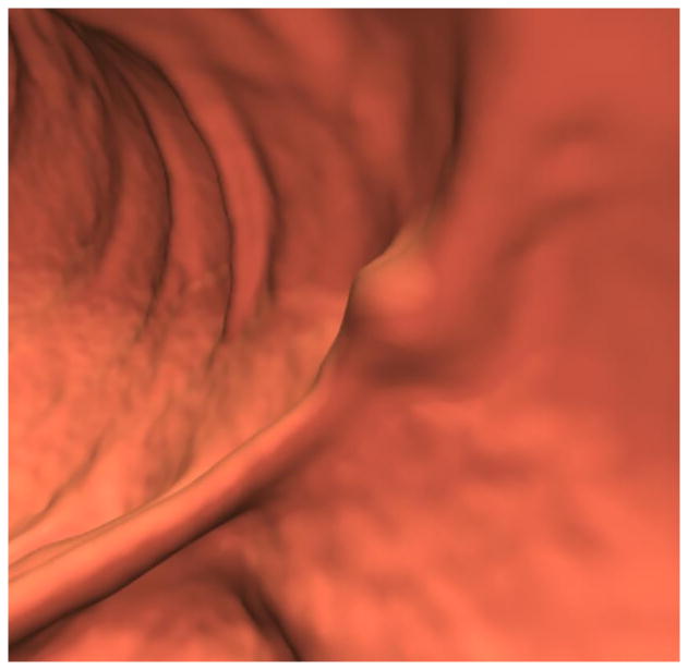
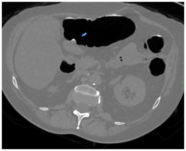
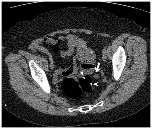
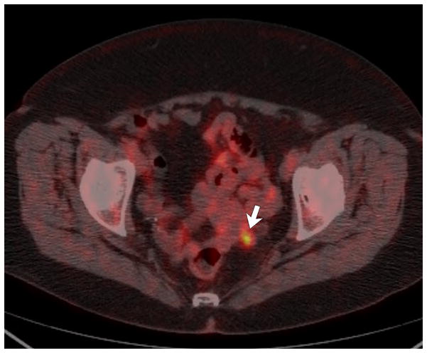
Results: None of the 201 patients had intraluminal anastomotic cancer recurrence or advanced neoplasia (or metachronous cancers). CT colonography detected extramural perianastomotic recurrence in 2 patients (1.0%); neither was detected at colonoscopy. Only 2 patients (1.0%; 2/201) were called positive at CT colonography for intraluminal anastomotic nondiminutive lesions (7- to 8-mm polyps), which were confirmed at colonoscopy but nonneoplastic at histopathology. At optical colonoscopy, the anastomosis was deemed abnormal and/or biopsied in 10.0% (20/201), yielding only 1 nondiminutive benign neoplasm (7-mm tubular adenoma).
Limitations: The lack of luminal cancer recurrence in our lower-risk cohort precludes assessment of sensitivity for detection, rendering the study underpowered in this regard. Potential cost savings of combined CT/CT colonography over the standard CT/colonoscopy approach were not assessed.
Conclusions: Relevant intraluminal anastomotic pathology appears to be very uncommon 1 year after colorectal cancer resection in lower-risk cohorts. Unlike colonoscopy, diagnostic contrast-enhanced CT colonography effectively evaluates both the intra- and extraluminal aspects of the anastomosis. See Video Abstract at http://links.lww.com/DCR/A471.
Figures


Similar articles
-
CT colonography for combined colonic and extracolonic surveillance after curative resection of colorectal cancer.Radiology. 2010 Dec;257(3):697-704. doi: 10.1148/radiol.10100385. Epub 2010 Sep 27. Radiology. 2010. PMID: 20876390
-
Adenomatous neoplasia: postsurgical incidence after normal preoperative CT colonography findings in the colon proximal to an occlusive cancer.Radiology. 2014 Oct;273(1):99-107. doi: 10.1148/radiol.14132844. Epub 2014 Jun 11. Radiology. 2014. PMID: 24918959
-
Post-surgical follow-up of colorectal cancer: role of contrast-enhanced CT colonography.Abdom Imaging. 2010 Dec;35(6):669-75. doi: 10.1007/s00261-009-9596-6. Abdom Imaging. 2010. PMID: 20033808
-
CT colonography for follow-up after surgery for colorectal cancer.AJR Am J Roentgenol. 2007 Aug;189(2):283-9. doi: 10.2214/AJR.07.2305. AJR Am J Roentgenol. 2007. PMID: 17646452 Review.
-
Imaging alternatives to colonoscopy: CT colonography and colon capsule. European Society of Gastrointestinal Endoscopy (ESGE) and European Society of Gastrointestinal and Abdominal Radiology (ESGAR) Guideline - Update 2020.Eur Radiol. 2021 May;31(5):2967-2982. doi: 10.1007/s00330-020-07413-4. Eur Radiol. 2021. PMID: 33104846 Review.
Cited by
-
Efficacy of endoscopic surveillance in the detection of local recurrence after radical rectal cancer surgery is limited? A retrospective study.World J Surg Oncol. 2021 Oct 21;19(1):308. doi: 10.1186/s12957-021-02413-0. World J Surg Oncol. 2021. PMID: 34670554 Free PMC article.
-
Computed Tomography Colonography vs Colonoscopy for Colorectal Cancer Surveillance After Surgery.Gastroenterology. 2018 Mar;154(4):927-934.e4. doi: 10.1053/j.gastro.2017.11.025. Epub 2017 Nov 22. Gastroenterology. 2018. PMID: 29174927 Free PMC article. Clinical Trial.
-
Reevaluating the Evidence for Intensive Postoperative Extracolonic Surveillance for Nonmetastatic Colorectal Cancer.Value Health. 2022 Jan;25(1):36-46. doi: 10.1016/j.jval.2021.07.017. Epub 2021 Oct 13. Value Health. 2022. PMID: 35031098 Free PMC article.
-
British Society of Gastroenterology/Association of Coloproctology of Great Britain and Ireland/Public Health England post-polypectomy and post-colorectal cancer resection surveillance guidelines.Gut. 2020 Feb;69(2):201-223. doi: 10.1136/gutjnl-2019-319858. Epub 2019 Nov 27. Gut. 2020. PMID: 31776230 Free PMC article.
References
-
- Kahi CJ, Boland CR, Dominitz JA, et al. Colonoscopy Surveillance After Colorectal Cancer Resection: Recommendations of the US Multi-Society Task Force on Colorectal Cancer. Gastroenterology. 2016;150:758–68. - PubMed
-
- Rex DK, Kahi CJ, Levin B, et al. Guidelines for colonoscopy surveillance after cancer resection: a consensus update by the American Cancer Society and US Multi-Society Task Force on Colorectal Cancer. CA Cancer J Clin. 2006;56:160–167. - PubMed
-
- Kim DH, Pickhardt PJ, Taylor AJ. Characteristics of advanced adenomas detected at CT colonographic screening: Implications for appropriate polyp size thresholds for polypectomy versus surveillance. AJR Am J Roentgenol. 2007;188:940–944. - PubMed
-
- Choi YJ, Park SH, Lee SS, et al. CT colonography for follow-up after surgery for colorectal cancer. AJR Am J Roentgenol. 2007;189:283–289. - PubMed
Publication types
MeSH terms
Grants and funding
LinkOut - more resources
Full Text Sources
Other Literature Sources
Medical
