

Differential Proteome Analysis of Extracellular Vesicles from Breast Cancer Cell Lines by Chaperone Affinity Enrichment
- PMID: 28991197
- PMCID: PMC5748560
- DOI: 10.3390/proteomes5040025
Differential Proteome Analysis of Extracellular Vesicles from Breast Cancer Cell Lines by Chaperone Affinity Enrichment
Abstract
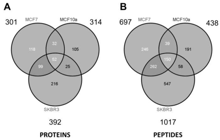
The complexity of human tissue fluid precludes timely identification of cancer biomarkers by immunoassay or mass spectrometry. An increasingly attractive strategy is to primarily enrich extracellular vesicles (EVs) released from cancer cells in an accelerated manner compared to normal cells. The Vn96 peptide was herein employed to recover a subset of EVs released into the media from cellular models of breast cancer. Vn96 has affinity for heat shock proteins (HSPs) decorating the surface of EVs. Reflecting their cells of origin, cancer EVs displayed discrete differences from those of normal phenotype. GELFrEE LC/MS identified an extensive proteome from all three sources of EVs, the vast majority having been previously reported in the ExoCarta database. Pathway analysis of the Vn96-affinity proteome unequivocally distinguished EVs from tumorigenic cell lines (SKBR3 and MCF-7) relative to a non-tumorigenic source (MCF-10a), particularly with regard to altered metabolic enzymes, signaling, and chaperone proteins. The protein data sets provide valuable information from material shed by cultured cells. It is probable that a vast amount of biomarker identities may be collected from established and primary cell cultures using the approaches described here.
Keywords: GELFrEE; Vn96; breast cancer cell lines; extracellular vesicles; glycolysis; heat shock proteins.
Conflict of interest statement
The authors declare no conflict of interest.
Figures
Similar articles
-
Proteome profiling of extracellular vesicles captured with the affinity peptide Vn96: comparison of Laemmli and TRIzol© protein-extraction methods.J Extracell Vesicles. 2018 Feb 26;7(1):1438727. doi: 10.1080/20013078.2018.1438727. eCollection 2018. J Extracell Vesicles. 2018. PMID: 29511462 Free PMC article.
-
Cell Stress Induced Stressome Release Including Damaged Membrane Vesicles and Extracellular HSP90 by Prostate Cancer Cells.Cells. 2020 Mar 19;9(3):755. doi: 10.3390/cells9030755. Cells. 2020. PMID: 32204513 Free PMC article.
-
Feasibility of urinary extracellular vesicle proteome profiling using a robust and simple, clinically applicable isolation method.J Extracell Vesicles. 2017 Apr 28;6(1):1313091. doi: 10.1080/20013078.2017.1313091. eCollection 2017. J Extracell Vesicles. 2017. PMID: 28717416 Free PMC article.
-
Proteomics for comprehensive characterization of extracellular vesicles in neurodegenerative disease.Exp Neurol. 2022 Sep;355:114149. doi: 10.1016/j.expneurol.2022.114149. Epub 2022 Jun 19. Exp Neurol. 2022. PMID: 35732219 Review.
-
Chromatography and its hyphenation to mass spectrometry for extracellular vesicle analysis.J Chromatogr A. 2016 Mar 25;1439:26-41. doi: 10.1016/j.chroma.2016.01.017. Epub 2016 Jan 11. J Chromatogr A. 2016. PMID: 26830636 Review.
Cited by
-
Nano-Bio Interactions of Extracellular Vesicles with Gold Nanoislands for Early Cancer Diagnosis.Research (Wash D C). 2018 Oct 9;2018:3917986. doi: 10.1155/2018/3917986. eCollection 2018. Research (Wash D C). 2018. PMID: 31549028 Free PMC article.
-
Rapid Activation of Neuroinflammation in Stroke: Plasma and Extracellular Vesicles Obtained on a Mobile Stroke Unit.Stroke. 2023 Mar;54(3):e52-e57. doi: 10.1161/STROKEAHA.122.041422. Epub 2023 Feb 2. Stroke. 2023. PMID: 36727508 Free PMC article.
-
Molecular interactions at the surface of extracellular vesicles.Semin Immunopathol. 2018 Sep;40(5):453-464. doi: 10.1007/s00281-018-0682-0. Epub 2018 Apr 16. Semin Immunopathol. 2018. PMID: 29663027 Free PMC article. Review.
-
Investigating the consistency of extracellular vesicle production from breast cancer subtypes using CELLine adherent bioreactors.J Extracell Biol. 2022 Sep 22;1(9):e60. doi: 10.1002/jex2.60. eCollection 2022 Sep. J Extracell Biol. 2022. PMID: 38938775 Free PMC article.
-
Proteome profiling of extracellular vesicles captured with the affinity peptide Vn96: comparison of Laemmli and TRIzol© protein-extraction methods.J Extracell Vesicles. 2018 Feb 26;7(1):1438727. doi: 10.1080/20013078.2018.1438727. eCollection 2018. J Extracell Vesicles. 2018. PMID: 29511462 Free PMC article.
References
-
- Americal Cancer Society. [(accessed on 25 July 2017)]; Available online: https://www.cancer.org/cancer/breast-cancer/understanding-a-breast-cance....
-
- Lehman C.D., Isaacs C., Schnall M.D., Pisano E.D., Ascher S.M., Weatherall P.T., Bluemke D.A., Owen D.J., Marcom P.K., Armstrong D.K., et al. Cancer yield of mammography, MR, and US in high-risk women: Prospective multi-institution breast cancer screening study. Radiology. 2007;244:381–388. doi: 10.1148/radiol.2442060461. - DOI - PubMed
-
- Berg W.A., Zhang Z., Lehrer D., Jong R.A., Pisano E.D., Barr R.G., Böhm-Vélez M., Mahoney M.C., Evans W.P., 3rd, Larsen L.H., et al. Detection of breast cancer with addition of annual screening ultrasound or a single screening MRI to mammography in women with elevated breast cancer risk. JAMA. 2012;307:1394–1404. doi: 10.1001/jama.2012.388. - DOI - PMC - PubMed
LinkOut - more resources
Full Text Sources
Other Literature Sources
