. 2017 Nov;49(11):1593-1601.
doi: 10.1038/ng.3970. Epub 2017 Oct 9.

Contribution of rare inherited and de novo variants in 2,871 congenital heart disease probands

Affiliations

Contribution of rare inherited and de novo variants in 2,871 congenital heart disease probands

Sheng Chih Jin et al. Nat Genet. 2017 Nov.

Abstract

Congenital heart disease (CHD) is the leading cause of mortality from birth defects. Here, exome sequencing of a single cohort of 2,871 CHD probands, including 2,645 parent-offspring trios, implicated rare inherited mutations in 1.8%, including a recessive founder mutation in GDF1 accounting for ∼5% of severe CHD in Ashkenazim, recessive genotypes in MYH6 accounting for ∼11% of Shone complex, and dominant FLT4 mutations accounting for 2.3% of Tetralogy of Fallot. De novo mutations (DNMs) accounted for 8% of cases, including ∼3% of isolated CHD patients and ∼28% with both neurodevelopmental and extra-cardiac congenital anomalies. Seven genes surpassed thresholds for genome-wide significance, and 12 genes not previously implicated in CHD had >70% probability of being disease related. DNMs in ∼440 genes were inferred to contribute to CHD. Striking overlap between genes with damaging DNMs in probands with CHD and autism was also found.


Conflict of interest statement

COMPETING FINANCIAL INTERESTS

None

Figures

Figure 1
Figure 1. Quantile-quantile plots comparing observed versus expected P-values for recessive genotypes in each gene in cases and controls
Recessive genotypes (RGs) shown include LoF, D-Mis, and non frameshift insertion/deletions. The expected number of RGs in each gene was calculated from the total number of observed RGs as described in Methods. The significance of the difference between the observed and expected number of RGs was calculated using a one-sided binomial test. (a). Quantile-quantile (Q-Q) plot in cases. (b). Q-Q plot in controls. While the observed values closely conform to expected values in controls, two genes, GDF1 and MYH6, show a significantly increased burden of RGs in cases and survive the multiple-testing correction threshold.
Figure 2
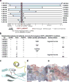
Figure 2. Phenotypes and shared haplotypes among homozygotes for GDF1-p.Met364Thr
(a). Extent of homozygous SNPs flanking homozygous GDF1-p.Met364Thr genotypes. A 5.9 Mb segment of chromosome 19 extending across the location of the homozygous GDF1-p.Met364Thr mutation (denoted by red square) in each unrelated subject is depicted. At the bottom, tick marks indicate location of all SNPs found by exome sequencing among Ashkenazim in cases. Known SNPs are shown via their rs identifiers. Allele frequencies of novel SNPs are indicated by asterisks. The closest heterozygous SNP to either side of the GDF1-p.Met364Thr in each subject is shown as a white square; all SNPs between these two heterozygous SNPs, encompassed by the light blue bar, are homozygous for the same allele seen in other subjects, consistent with the p.Met364Thr variant being identical by descent among all subjects. The length of each homozygous segment is indicated at the right of the panel. The maximum length of the homozygous segment shared by all subjects is 234 kb (shown as grey vertical bar), consistent with the mutation having been introduced into a shared ancestor many generations ago. (b). Cardiac and extracardiac phenotypes of GDF1-p.Met364Thr homozygotes. Present phenotypes are denoted with ‘+’, those absent with ‘−’, and those unavailable for testing with ‘NA’ (c). Ribbon diagram of part of GDF1 homodimer containing p.Met364. The hydrophobic helix from one subunit (yellow) sits above p.Met364 on the other subunit (blue). (d). Space filling model of the segment of GDF1 containing the wild-type p.Met364 showing surface electrostatic charge (blue=positive, red=negative). (e). Surface electrostatic charge of the segment containing mutant p.Thr364. Compared to wild-type, the mutant peptide shows a more negatively charged cavity.
Figure 3
Figure 3. FLT4 loss-of-function mutations in Tetralogy of Fallot
(a). Pedigrees of 10 CHD kindreds with rare FLT4 loss-of-function (LoF) mutations are shown. Subjects with and without CHD are shown as filled and unfilled symbols, respectively. Each kindred ID number is shown along with the FLT4 genotype of each subject and CHD phenotype of affected subjects. (b) Diagram of FLT4 protein is shown with seven immunoglobulin domains (Ig) and a kinase domain. The top panel shows LoF mutations associated with Tetralogy-type CHD, whereas the bottom panel displays missense mutations associated with the Milroy disease (Hereditary Lymphedema).
Figure 4
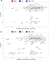
Figure 4. Chromatin modification genes and genes with multiple damaging de novo mutations are enriched for high expression in developing heart and intolerance to loss-of-function mutation
(a) Enrichment of damaging mutations in chromatin modifiers in genes highly expressed in developing heart and intolerant to loss-of-function (LoF) mutation. X axis (0–100) denotes the percentile rank of heart expression in developing mouse heart at E14.5, and y axis (0–1.0) denotes intolerance to LoF mutation (pLI) in the ExAC database. (b) 66 genes with 2 or more damaging de novo mutations are plotted. Multihit genes are highly enriched (N=31) for genes that are highly expressed in developing heart and intolerant to LoF mutation (pLI ≥ 0.99).

Comment in

Similar articles

  • Genomic analysis of 11,555 probands identifies 60 dominant congenital heart disease genes.
    Sierant MC, Jin SC, Bilguvar K, Morton SU, Dong W, Jiang W, Lu Z, Li B, López-Giráldez F, Tikhonova I, Zeng X, Lu Q, Choi J, Zhang J, Nelson-Williams C, Knight JR, Zhao H, Cao J, Mane S, Sedore SC, Gruber PJ, Lek M, Goldmuntz E, Deanfield J, Giardini A, Mital S, Russell M, Gaynor JW, King E, Wagner M, Srivastava D, Shen Y, Bernstein D, Porter GA Jr, Newburger JW, Seidman JG, Roberts AE, Yandell M, Yost HJ, Tristani-Firouzi M, Kim R, Chung WK, Gelb BD, Seidman CE, Brueckner M, Lifton RP. Sierant MC, et al. Proc Natl Acad Sci U S A. 2025 Apr;122(13):e2420343122. doi: 10.1073/pnas.2420343122. Epub 2025 Mar 24. Proc Natl Acad Sci U S A. 2025. PMID: 40127276 Free PMC article.
  • Recessive genetic contribution to congenital heart disease in 5,424 probands.
    Dong W, Jin SC, Sierant MC, Lu Z, Li B, Lu Q, Morton SU, Zhang J, López-Giráldez F, Nelson-Williams C, Knight JR, Zhao H, Cao J, Mane S, Gruber PJ, Lek M, Goldmuntz E, Deanfield J, Giardini A, Mital S, Russell M, Gaynor JW, Cnota JF, Wagner M, Srivastava D, Bernstein D, Porter GA Jr, Newburger J, Roberts AE, Yandell M, Yost HJ, Tristani-Firouzi M, Kim R, Seidman J, Chung WK, Gelb BD, Seidman CE, Lifton RP, Brueckner M. Dong W, et al. Proc Natl Acad Sci U S A. 2025 Mar 11;122(10):e2419992122. doi: 10.1073/pnas.2419992122. Epub 2025 Mar 3. Proc Natl Acad Sci U S A. 2025. PMID: 40030011 Free PMC article.
  • Utilization of Whole Exome Sequencing to Identify Causative Mutations in Familial Congenital Heart Disease.
    LaHaye S, Corsmeier D, Basu M, Bowman JL, Fitzgerald-Butt S, Zender G, Bosse K, McBride KL, White P, Garg V. LaHaye S, et al. Circ Cardiovasc Genet. 2016 Aug;9(4):320-9. doi: 10.1161/CIRCGENETICS.115.001324. Epub 2016 Jul 14. Circ Cardiovasc Genet. 2016. PMID: 27418595 Free PMC article.
  • The genetics of isolated congenital heart disease.
    Nees SN, Chung WK. Nees SN, et al. Am J Med Genet C Semin Med Genet. 2020 Mar;184(1):97-106. doi: 10.1002/ajmg.c.31763. Epub 2019 Dec 26. Am J Med Genet C Semin Med Genet. 2020. PMID: 31876989 Free PMC article. Review.
  • Genetics of congenital heart disease: the contribution of the noncoding regulatory genome.
    Postma AV, Bezzina CR, Christoffels VM. Postma AV, et al. J Hum Genet. 2016 Jan;61(1):13-9. doi: 10.1038/jhg.2015.98. Epub 2015 Jul 30. J Hum Genet. 2016. PMID: 26223183 Review.

Cited by

References

    1. van der Linde D, et al. Birth prevalence of congenital heart disease worldwide: a systematic review and meta-analysis. J Am Coll Cardiol. 2011;58:2241–7. - PubMed
    1. Egbe A, Lee S, Ho D, Uppu S, Srivastava S. Prevalence of congenital anomalies in newborns with congenital heart disease diagnosis. Ann Pediatr Cardiol. 2014;7:86–91. - PMC - PubMed
    1. Marino BS, et al. Neurodevelopmental outcomes in children with congenital heart disease: evaluation and management: a scientific statement from the American Heart Association. Circulation. 2012;126:1143–72. - PubMed
    1. Soemedi R, et al. Contribution of global rare copy-number variants to the risk of sporadic congenital heart disease. Am J Hum Genet. 2012;91:489–501. - PMC - PubMed
    1. Glessner JT, et al. Increased frequency of de novo copy number variants in congenital heart disease by integrative analysis of single nucleotide polymorphism array and exome sequence data. Circ Res. 2014;115:884–96. - PMC - PubMed

MeSH terms