. 2017 Nov;24(11):986-992.
doi: 10.1038/nsmb.3484. Epub 2017 Oct 9.

Structural basis for GABAA receptor potentiation by neurosteroids

Affiliations

Structural basis for GABAA receptor potentiation by neurosteroids

Paul S Miller et al. Nat Struct Mol Biol. 2017 Nov.

Abstract

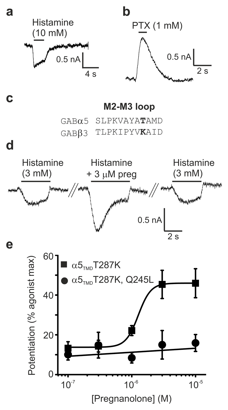
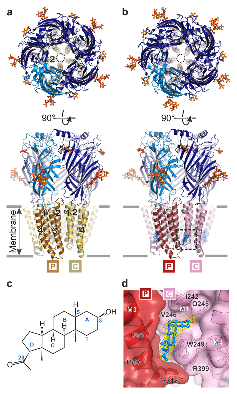
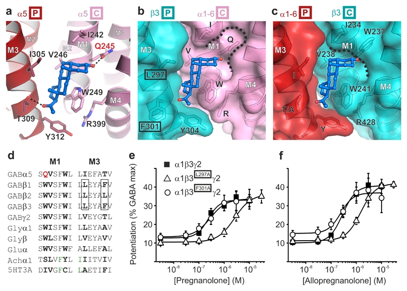
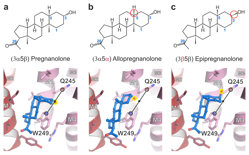
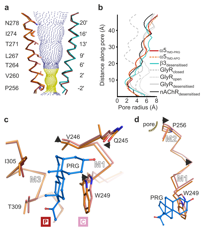
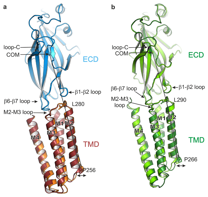
Type A γ-aminobutyric acid receptors (GABAARs) are the principal mediators of inhibitory neurotransmission in the human brain. Endogenous neurosteroids interact with GABAARs to regulate acute and chronic anxiety and are potent sedative, analgesic, anticonvulsant and anesthetic agents. Their mode of binding and mechanism of receptor potentiation, however, remain unknown. Here we report crystal structures of a chimeric GABAAR construct in apo and pregnanolone-bound states. The neurosteroid-binding site is mechanically coupled to the helices lining the ion channel pore and modulates the desensitization-gate conformation. We demonstrate that the equivalent site is responsible for physiological, heteromeric GABAAR potentiation and explain the contrasting modulatory properties of 3a versus 3b neurosteroid epimers. These results illustrate how peripheral lipid ligands can regulate the desensitization gate of GABAARs, a process of broad relevance to pentameric ligand-gated ion channels.


Conflict of interest statement

Competing Financial Interests

The authors declare no competing financial interests.

Figures

Figure 1
Figure 1
α5TMD forms a functional gating unit potentiated by pregnanolone at the Gln245 site. (a) HEK-293T whole-cell patch-clamp recording of the agonist histamine activating an inward current through α5TMD, that subsequently desensitizes. (b) The classical GABAAR channel blocker picrotoxin blocks a spontaneous inward leak current generated by α5TMD. (c) Sequence alignment of the M2-M3 loop from GABAAR α5 and β3 subunits, highlighting in bold the T287K substitution. (d) Raw whole-cell patch-clamp EC10 histamine responses from HEK cells transfected with α5TMDT287K, before and after co-application with 3 μM pregnanolone. (e) Pregnanolone dose-response curve for potentiation of histamine EC10-15 responses in α5TMD T287K, and the ablation of potentiation upon introduction of a Q245L mutation in the M1 helix. Each data point represents mean ± s.e.m. of n = 6 experiments, each measurement being from different cells.
Figure 2
Figure 2
Architecture of GABAAR α5TMD. (a) and (b) Top-down and side-on views of the α5TMD pentamer in apo and pregnanolone-bound forms, respectively. Subunits each comprise the β3 extracellular domain (dark and light blue) and α5 transmembrane domain (α-helices M1-M4 shown in beige/yellow or red/pink). In top-down views, the pore is indicated by a black circle, lined by five M2 helices (one per subunit). Side-on views highlight the helical organisation (M1-M4, numbered 1 to 4 per subunit, where visible) in (a), and the presence of pregnanolone in (b) (blue stick and space-fill, inside dashed box). N-linked glycans are shown as spheres coloured by atom type (carbon atoms in orange, oxygen atoms in red). The principal (P) and complementary (C) subunits, for the visible inter-subunit interface, are indicated by boxed labels. (c) Structural formula of pregnanolone with rings labelled A to D. Carbon atoms mentioned in the text are numbered. (d) Close-up of pregnanolone ("ball-and-stick" representation) bound to α5TMD (cartoon and surface representation). The 2Fo-Fc electron density map surrounding the ligand is contoured at 1.3σ (yellow mesh).
Figure 3
Figure 3
The neurosteroid potentiation site. (a) Pregnanolone (ball-and-stick representation, carbon atoms in blue, oxygen atoms in red) bound to an inter-subunit site between M3 residues on the principal face of one subunit (P in box) and M1 residues on the complementary face of the next (C in box). Putative hydrogen bonds between the pregnanolone 3α-hydroxyl and the Gln245 amide (2.4 Å) and C20 ketone and Thr309 hydroxyl (3.0 Å) are indicated by dashed lines. (b) and (c) Modelled heteromeric βPC and αPC interfaces respectively, in cartoon and surface representation, in which the relevant α5TMD face is substituted by the equivalent β3 subunit structure (PDB ID: 4COF). Note that α5 residues in these two panels are labelled by letters only, without numbers, to reflect their conservation across the α subunits (α1-6) within this site. Pregnanolone is well accommodated in the heteromeric βPC site (b) but not in the αCP site (c) due to the replacement of α M1 Gln with the β3 M1 Trp237, which closes the top of the pocket (dashed line). (d) Sequence alignment of the M1 and M3 motifs containing the neurosteroid binding site residues (bold) in the GABAAR α5 subunit and equivalent regions in other human pLGIC superfamily members. M1 Gln245, unique to GABAAR α-subunits, is highlighted in red. GABAAR β-subunit residues contributed from the complementary face in heteromeric receptors are boxed. (e) Pregnanolone and (f) Allopregnanolone dose-response curves for potentiation of GABA EC10-15 responses. Alanine substitution of β3 principal face residues Leu297, but not Phe301, reduces neurosteroid sensitivity. These residues are highlighted by black boxes in b. Each data point represents mean ± s.e.m. Pregnanolone EC50 n number: EC50: WT n = 13, L297A n = 11, F301A n = 8. Allopregnanolone EC50: WT n = 5, L297A n = 9, F301A n = 4. Each n EC50 measurement is from different cells.
Figure 4
Figure 4
Computational docking of pregnanolone and related neurosteroids. Structural formulae and α5TMD structures showing binding modes of computationally docked (a) pregnanolone, (b) allopregnanolone and (c) epipregnanolone. Stereoisomeric differences from pregnanolone are indicated in formulae by red circles. In the 3D models, the critical C3 hydroxyl group is highlighted by a yellow circle. Potentiators possess a 3α-hydroxyl which is computationally docked equidistantly between the Gln245 εO Gln and Trp249 εN (3.5 Å for pregnanolone; 3.6 Å for allopregnanolone; indicated by a black line) regardless of whether the rings assume a half-chair or flat geometry. The C3 hydroxyl of 3β pregnanolone can only share a putative hydrogen bond with the Gln245 amide (2.8 Å distance).
Figure 5
Figure 5
Pore conformation and neurosteroid induced motions in α5TMD. (a) View of two opposing pore-lining M2 helices, the other three being removed for clarity. The pregnanolone-bound α5TMD state (ruby) is shown on both sides. For comparison, overlays of α5TMD apo state (on the left side, orange), and of GABAAR-β3 (on the right side, teal) M2 helices are shown. Blue and yellow spheres define pregnanolone-bound α5TMD pore radii greater than or less than 3.2 Å respectively (radius of a hydrated Cl- ion). (b) A plot of pore radii for α5TMD structures compared to GABAAR-β3 in the desensitised state (PDB ID: 4COF), the glycine receptor in the resting closed, open and desensitised states (PDB IDs: 5JAD, 5JAE and 5JAF), and α4β2 nAChR (PDB ID: 5KXI). (c) Superposition of apo (orange) versus pregnanolone-bound (ruby) α5TMD principal faces reveals pregnanolone-induced motions on the complementary face (pink) of the neurosteroid pocket (indicated by short black arrows). (d) The same superposition as in c, zooming in on the pregnanolone-induced motions along the M1-M2 linker of a complementary face subunit, which impact on the Pro256 desensitization gate.
Figure 6
Figure 6
TMD motions relative to the ECD. (a) Side-on view of a single subunit (B chains), from apo α5TMD (orange TMD) and pregnanolone-bound α5TMD (ruby TMD), superposed by their ECDs (blue). The ECD conformations are very similar (RMSD of 0.19 Å over 204 equivalent Cα positions). However, the lower half of the pregnanolone-bound TMD flexes such that the desensitisation gate at Pro256 swings outwards from the pore (indicated by double-headed arrow). The angles measured between the ECD centre of mass (COM) and the Cα atoms of Leu280 at the top of M2 and Pro256 at the bottom of M2 are 162.5° (apo α5TMD) and 164.4° (pregnanolone-bound α5TMD), respectively, i.e. a 1.9° swing. (b) Equivalent view and superposition between open GlyR (light green, PDB ID: 3JAE) and desensitized (or partially open) GlyR (dark green, PDB ID: 3JAF). The narrowest pore diameter is at -2' (Pro266) position in both cases (8.8 Å and 5 Å, respectively). The GlyR ECD conformations are also very similar (RMSD of 0.31 Å over 210 equivalent Cα positions in chains B). As observed in α5TMD, relative to the superposed ECDs the lower half of the GlyR TMD flexes such that the desensitization gate at Pro266 is displaced outwards from the pore to expand and open the channel. The angles measured between the ECD COM and the Cα atoms of Leu290 at the top of M2 and Pro266 at the bottom of M2 are 161.4° (desensitized GlyR) and 164.1° (open GlyR), respectively, i.e. a 2.7° swing.

Similar articles

Cited by

References

    1. Nemecz A, Prevost MS, Menny A, Corringer PJ. Emerging Molecular Mechanisms of Signal Transduction in Pentameric Ligand-Gated Ion Channels. Neuron. 2016;90:452–70. - PubMed
    1. Miller PS, Smart TG. Binding, activation and modulation of Cys-loop receptors. Trends Pharmacol Sci. 2010;31:161–74. - PubMed
    1. Hosie AM, Wilkins ME, da Silva HM, Smart TG. Endogenous neurosteroids regulate GABAA receptors through two discrete transmembrane sites. Nature. 2006;444:486–9. - PubMed
    1. Majewska MD, Harrison NL, Schwartz RD, Barker JL, Paul SM. Steroid hormone metabolites are barbiturate-like modulators of the GABA receptor. Science. 1986;232:1004–7. - PubMed
    1. Sarkar J, Wakefield S, MacKenzie G, Moss SJ, Maguire J. Neurosteroidogenesis is required for the physiological response to stress: role of neurosteroid-sensitive GABAA receptors. J Neurosci. 2011;31:18198–210. - PMC - PubMed