. 2017 Nov 1;57(5):1071-1081.
doi: 10.1093/icb/icx116.

A Complex Lens for a Complex Eye

Affiliations

A Complex Lens for a Complex Eye

Aaron L Stahl et al. Integr Comp Biol. .

Abstract

A key innovation for high resolution eyes is a sophisticated lens that precisely focuses light onto photoreceptors. The eyes of holometabolous larvae range from very simple eyes that merely detect light to eyes that are capable of high spatial resolution. Particularly interesting are the bifocal lenses of Thermonectus marmoratus larvae, which differentially focus light on spectrally-distinct retinas. While functional aspects of insect lenses have been relatively well studied, little work has explored their molecular makeup, especially in regard to more complex eye types. To investigate this question, we took a transcriptomic and proteomic approach to identify the major proteins contributing to the principal bifocal lenses of T. marmoratus larvae. Mass spectrometry revealed 10 major lens proteins. Six of these share sequence homology with cuticular proteins, a large class of proteins that are also major components of corneal lenses from adult compound eyes of Drosophila melanogaster and Anopheles gambiae. Two proteins were identified as house-keeping genes and the final two lack any sequence homologies to known genes. Overall the composition seems to follow a pattern of co-opting transparent and optically dense proteins, similar to what has been described for other animal lenses. To identify cells responsible for the secretion of specific lens proteins, we performed in situ hybridization studies and found some expression differences between distal and proximal corneagenous cells. Since the distal cells likely give rise to the periphery and the proximal cells to the center of the lens, our findings highlight a possible mechanism for establishing structural differences that are in line with the bifocal nature of these lenses. A better understanding of lens composition provides insights into the evolution of proper focusing, which is an important step in the transition between low-resolution and high-resolution eyes.


Figures

Fig. 1
Fig. 1
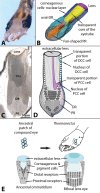
(A) The larvae of the sunburst diving beetle (Thermonectus marmoratus) are visually guided predators with six eyes on each side of the head, two of which are especially prominent. (B) A three-dimensional reconstruction of a principal eye showing its major components. They include corneagenous cells and the distal (DR) and proximal retinas (PR) (striped portions represent rhabdomes). The latter is fan-shaped, so that only two photoreceptor cells are visible in sagittal sections (C). The bifocal lens is needed to properly focus images on each retina. (C) A histological section of one of the principal eyes shows the cellular components of the four (two/side) principal larval eyes. (D) A schematic illustration of a sagittal section further illustrates its cellular composition. Note that the extracellular lens is positioned on top of the transparent portions of the corneagenous cells, which are known to be the source of lens secretion during development (Stecher et al. 2016). These cells are organized so that distal corneagenous cells (DCCs; see example cell in blue) underlie the lens’ periphery, whereas proximal corneagenous cells (PCCs; see example cell in purple) are in contact with the center of the lens. (E) Evidence suggests that each of the T. marmoratus eyes has evolved from a compound-eye ommatidial like ancestor (after Buschbeck 2014).
Fig. 2
Fig. 2
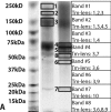
SDS–PAGE gel showing proteins extracted from T. marmoratus lenses and identified using mass spectrophotometry. Ten major proteins within eight bands were found. The left lane represents the molecular weight marker. The lens extract was loaded in two lanes on the right. Boxes indicate individual bands that were excised and subjected to a MALDI-TOF/TOF analysis.
Fig. 3
Fig. 3
This figure illustrates in situ labeling of mRNAs for lens proteins that are expressed at similar levels in distal (DCC) and proximal (PCC) cone cells. For Tm-lens3 and Tm-lens1, staining is also observed in the transparent portions of the eye tube. (A) Schematic representation of the expression patterns of these lens protein-encoding genes. The striped shading indicates that some, but not all genes analyzed led to staining in that area. (B) Tm-lens3 in situ labeling is very prominent in the entire eye tube. (C) Tm-lens1 in situ labeling overlaps with that of Tm-lens3, but staining is less intense. (D) Tm-lens4 in situ labeling is also observed in distal and PCCs, but not within the core of the eye tube. (E) A sense control for Tm-lens3 shows light staining artifacts in the distal retina (DR) when stained overnight (∼15 h). Stainings for B–D were <5 h.
Fig. 4
Fig. 4
In situ labeling of mRNAs for lens proteins that are expressed exclusively or more strongly in the DCCs. (A) Schematic representation of the expression patterns of these lens genes. The striped shading indicates that some, but not all of the genes are expressed in that area. The darker distal areas indicate more intense staining. These genes have in common that they are most strongly expressed in the DCCs. (B) Tm-lens10 in situ labeling indicates expression only in the most DCCs. (C and D) Tm-lens8 in situ labeling can be more (C) or less (D) restricted to distal cells. (E) Tm-lens2 generally showed low levels of in situ labeling, but was most strongly detected in DCCs.
Fig. 5
Fig. 5
In situ labeling of mRNAs for lens proteins that are expressed in all corneagenous cells, but with slightly more extensive staining in the proximal (vs. distal) corneagenous cells. (A) Schematic representation of the expression patterns of Tm-lens5-7 and 9. The striped shading indicates that not all four genes are expressed in that area. (B) Tm-lens9 in situ labeling is visible in all corneagenous cells, with slightly more extensive staining in the proximal, relative to distal, corneagenous cells. (C) T.m-lens5 in situ labeling shows similar, but weaker staining patterns as Tm-lens9. (D) Tm-lens7 shows labeling in the center portion of the most PCCs. (E) Tm-lens6 in situ labeling is similar to Tm-lens7 but with additional staining in distal retinula cells.

Similar articles

Cited by

References

    1. Altschul SF, Gish W, Miller W, Myers EW, Lipman DJ.. 1990. Basic local alignment search tool. J Mol Biol 215:403–10. - PubMed
    1. Andersen SO, Rafn K, Roepstorff P.. 1997. Sequence studies of proteins from larval and pupal cuticle of the yellow meal worm, Tenebrio molitor. Insect Biochem Mol Biol 27:121–31. - PubMed
    1. Blanco J, Girard F, Kamachi Y, Kondoh H, Gehring WJ.. 2005. Functional analysis of the chicken delta 1-crystallin enhancer activity in Drosophila reveals remarkable evolutionary conservation between chicken and fly. Development 132:1895–905. - PubMed
    1. Bland K, Revetta N, Stowasser A, Buschbeck EK. 2014. Unilateral range finding in diving beetle larvae. J Exp Biol 217:327–30. - PubMed
    1. Blest AD, Land MF.. 1977. Physiological optics of Dinopis subrufus L. Koch: a fish-lens in a spider. Proc R Soc Lond B Biol Sci 196:197–222. - PubMed

Publication types

LinkOut - more resources