. 2017 Oct 13;68(17):4765-4774.
doi: 10.1093/jxb/erx307.

Plasma membrane-associated cation-binding protein 1-like protein negatively regulates intercellular movement of BaMV

Affiliations

Plasma membrane-associated cation-binding protein 1-like protein negatively regulates intercellular movement of BaMV

Ying-Ping Huang et al. J Exp Bot. .

Abstract

To establish a successful infection, a virus needs to replicate and move cell-to-cell efficiently. We investigated whether one of the genes upregulated in Nicotiana benthamiana after Bamboo mosaic virus (BaMV) inoculation was involved in regulating virus movement. We revealed the gene to be a plasma membrane-associated cation-binding protein 1-like protein, designated NbPCaP1L. The expression of NbPCaP1L in N. benthamiana was knocked down using Tobacco rattle virus-based gene silencing and consequently the accumulation of BaMV increased significantly to that of control plants. Further analysis indicated no significant difference in the accumulation of BaMV in NbPCaP1L knockdown and control protoplasts, suggesting NbPCaP1L may affect cell-to-cell movement of BaMV. Using a viral vector expressing green fluorescent protein in the knockdown plants, the mean area of viral focus, as determined by fluorescence, was found to be larger in NbPCaP1L knockdown plants. Orange fluorescence protein (OFP)-fused NbPCaP1L, NbPCaP1L-OFP, was expressed in N. benthamiana and reduced the accumulation of BaMV to 46%. To reveal the possible interaction of viral protein with NbPCaP1L, we performed yeast two-hybrid and co-immunoprecipitation experiments. The results indicated that NbPCaP1L interacted with BaMV replicase. The results also suggested that NbPCaP1L could trap the BaMV movement RNP complex via interaction with the viral replicase in the complex and so restricted viral cell-to-cell movement.

Keywords: Bamboo mosaic virus; NbPCaP1L; Nicotiana benthamiana; defense protein; positive-sense RNA virus; replicase; viral RNA movement.


Figures

Fig. 1.
Fig. 1.
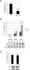
The relative accumulation of BaMV coat protein in NbPCaP1L knockdown plants and protoplasts. (A) The expression of NbPCaP1L mRNA was determined by real-time quantitative RT-PCR in Luc and NbPCaP1L knockdown leaves. (B) Western blot analysis of coat protein (CP) accumulation on the inoculated leaves. The accumulation of control plants was set to 100%. Data are mean ± standard deviation relative levels of BaMV CP from at least three independent experiments with at least three plants in each experiment. Representative western blot results of CP levels with triplicate results and rbcL as a control. (C) Western blot analysis of CP accumulation. The accumulation in control protoplasts at 24 h post inoculation (hpi) was set to 100%. Representative results are shown. Data are mean ± standard deviation relative levels of BaMV CP from at least three independent experiments. Luc, luciferase knockdown control plants or protoplasts; NbPCaP1L, NbPCaP1L knockdown plants or protoplasts; CP, BaMV coat protein; rbcL, RuBisCO large subunit (the loading control for normalization). **P<0.01, ***P<0.001 by Student’s t-test.
Fig. 2.
Fig. 2.
Cell-to-cell movement of BaMV in Luc and NbPCaP1L knockdown plants. (A) Fluorescent microscopy of the area of fluorescent foci in inoculated leaves of luciferase knockdown control (Luc) and NbPCaP1L knockdown (NbPCaP1L) plants after inoculation with the BaMV infectious plasmid carrying green fluorescent protein (GFP). Scale bar, 2 mm. (B) Statistical analysis. Data are mean ± standard deviation of 19 and 29 foci from Luc and NbPCaP1L knockdown plants, respectively. **P<0.01 by Student’s t-test.
Fig. 3.
Fig. 3.
The localization at the plasma membrane (PM) and cytoplasm of NbPCaP1L with OFP fused at the C-terminus and transiently expressed in N. benthamiana protoplasts. Cyan fluorescence protein (CFP) fused with a plasma membrane aquaporin AtPIP2A is transiently expressed in protoplasts and is used as the PM marker.
Fig. 4.
Fig. 4.
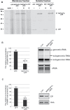
Effect of expression of NbPCaP1L on BaMV infection. (A) Western blot analysis of the fractionation of transiently expressed NbPCaP1L-OFP and OFP alone. Relative accumulation of BaMV CP (B) and RNAs (C) at 5 d post-inoculation (dpi) on inoculated leaves, with NbPCaP1L-OFP or OFP only (as a control) transiently expressed on the same leaves at 3 dpi. Data are mean ± standard error of at least three independent experiments. **P<0.01 by Student’s t-test.
Fig. 5.
Fig. 5.
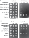
Interaction of NbPCaP1L with BaMV-encoded polypeptides in yeast cells. Yeast strain L40 co-transformed with the prep plasmid, pYES-TGBp1, -2, -3, or -CP, and bait plasmid pLEX-NbPCaP1L in (A) or prey plasmid pYES-NbPCaP1L and bait plasmid, pLEX-Capping, -RdRp, or -Helicase in (B). Positive control, yeast containing pLEX-Fos2 and pYES-Jun; negative control, yeast containing pHybLex/Zeo and pYESTrp2. Positive colonies were grown on Trp-/His-/Zeocin selection agar plates. The yeast concentrations with the dilution factor are indicated at the top of each panel.
Fig. 6.
Fig. 6.
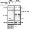
Co-immunoprecipitation (IP) assay to verify the interaction between BaMV replication protein and NbPCaP1L. Total proteins (input) were extracted from N. benthamiana leaves co-infiltrated with Agrobacteria containing the infectious BaMV/Rep-HA and OFP vector or NbPCaP1L-OFP. Equal amounts of input proteins were used for immunoblotting (IB) with antibodies against HA, OFP, and BaMV CP as indicated. rbcL, RuBisCo large subunit is used as the loading control. After IP with anti-HA antibody, the co-IP of Rep-HA, NbPCaP1L-OFP, and CP was detected using corresponding antibodies for IB.

Similar articles

Cited by

References

    1. Agre P, Bonhivers M, Borgnia MJ. 1998. The aquaporins, blueprints for cellular plumbing systems. The Journal of Biological Chemistry 273, 14659–14662. - PubMed
    1. Avisar D, Prokhnevsky AI, Dolja VV. 2008. Class VIII myosins are required for plasmodesmatal localization of a closterovirus Hsp70 homolog. Journal of Virology 82, 2836–2843. - PMC - PubMed
    1. Baurès I, Candresse T, Leveau A, Bendahmane A, Sturbois B. 2008. The Rx gene confers resistance to a range of potexviruses in transgenic Nicotiana plants. Molecular Plant-Microbe Interactions 21, 1154–1164. - PubMed
    1. Benitez-Alfonso Y, Faulkner C, Ritzenthaler C, Maule AJ. 2010. Plasmodesmata: gateways to local and systemic virus infection. Molecular Plant-Microbe Interactions 23, 1403–1412. - PubMed
    1. Chaumont F, Barrieu F, Jung R, Chrispeels MJ. 2000. Plasma membrane intrinsic proteins from maize cluster in two sequence subgroups with differential aquaporin activity. Plant Physiology 122, 1025–1034. - PMC - PubMed

Publication types