. 2018 Jan;70(1):88-97.
doi: 10.1002/art.40342. Epub 2017 Dec 1.

Visualization of Peripheral Neuron Sensitization in a Surgical Mouse Model of Osteoarthritis by In Vivo Calcium Imaging

Affiliations

Visualization of Peripheral Neuron Sensitization in a Surgical Mouse Model of Osteoarthritis by In Vivo Calcium Imaging

Rachel E Miller et al. Arthritis Rheumatol. 2018 Jan.

Abstract

Objective: To develop a method for analyzing sensory neuron responses to mechanical stimuli in vivo, and to evaluate whether these neuronal responses change after destabilization of the medial meniscus (DMM).

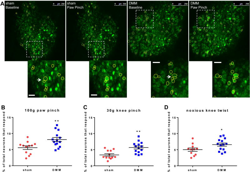
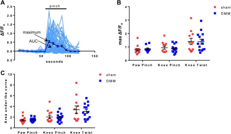
Methods: DMM or sham surgery was performed in 10-week-old male C57BL/6 wild-type or Pirt-GCaMP3+/- mice. All experiments were performed 8 weeks after surgery. Knee and hind paw hyperalgesia were assessed in wild-type mice. The retrograde label DiI was injected into the ipsilateral knee to quantify the number of knee-innervating neurons in the L4 dorsal root ganglion (DRG) in wild-type mice. In vivo calcium imaging was performed on the ipsilateral L4 DRG of Pirt-GCaMP3+/- mice as mechanical stimuli (paw pinch, knee pinch, or knee twist) were applied to the ipsilateral hind limb.

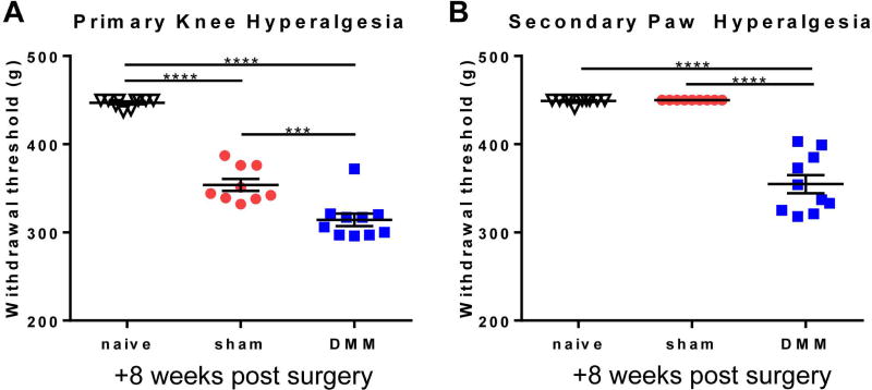
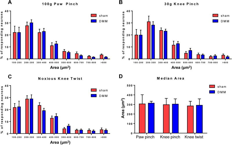
Results: Eight weeks after surgery, mice subjected to DMM had more hyperalgesia in the knee and hind paw compared to mice subjected to sham surgery. Intraarticular injection of DiI labeled similar numbers of neurons in the L4 DRG of mice subjected to sham surgery and mice subjected to DMM. Increased numbers of sensory neurons responded to all 3 mechanical stimuli in mice subjected to DMM, as assessed by in vivo calcium imaging. The majority of responses in mice subjected to sham surgery and mice subjected to DMM were in small to medium-sized neurons, consistent with the size of nociceptors. The magnitude of responses was similar between mice subjected to sham surgery and mice subjected to DMM.

Conclusion: Our findings indicate that increased numbers of small to medium-sized DRG neurons respond to mechanical stimuli 8 weeks after DMM surgery, suggesting that nociceptors have become sensitized by lowering the response threshold.


Figures

Figure 1
Figure 1
Wild-type mice that have undergone DMM surgery have increased (A) primary knee hyperalgesia and (B) secondary paw hyperalgesia 8 weeks after surgery compared to sham and age-matched naïve mice (***p<0.001, ****p<0.0001). mean±SEM.
Figure 2
Figure 2
Eight weeks after surgery, paw- and knee-directed stimuli cause increased numbers of L4 DRG neuron responses in DMM mice compared to sham mice. (A) Representative images depicting L4 DRG neurons at baseline and responding to a 100 g pinch of the ipsilateral hind paw. Scale bars = 250 µm. Insets show examples of responding neurons shown with the arrow. The arrow also identifies examples of neuron clusters. Scale bar in insets = 50 µm. (B) Percent of total neurons responding to 100 g ipsilateral paw pinch (# responding neurons / # total neurons in the field of view × 100) (sham: 5.6±0.6%; DMM: 8.1±0.6%). (C) Percent of total neurons responding to 30 g ipsilateral knee pinch (sham: 3.4±0.4%; DMM: 5.7±0.5%). (D) Percent of total neurons responding to noxious knee twist (sham: 5.0±0.5%; DMM: 6.6±0.5%). Each dot shows the percent of neurons that responded to the stimulus in one mouse L4 DRG. *p<0.05, **p<0.01. mean±SEM.
Figure 3
Figure 3
The size of responding neurons is similar in DMM and sham mice. (A–C) Relative frequency distributions of the areas of responding neurons in sham and DMM mice to (A) 100 g paw pinch (interaction of size and treatment (sham/DMM): p=0.9888), (B) 30 g knee pinch (interaction of size and treatment: p=0.9912), and (C) noxious knee twist (interaction of size and treatment: p=0.9474). mean±SEM. (D) Median area of responding cells. Sham vs. DMM, paw pinch: p=0.6770; knee pinch: p=0.9798; knee twist: p=0.8089. median±IQR.
Figure 4
Figure 4
The spatial organization of responding neurons is similar in DMM and sham mice. (A) The percentage of responding neurons that were coupled in each DRG (# responding neurons touching at least one other responding neuron / total # responding neurons × 100). Sham vs. DMM, paw pinch: p=0.6222; knee pinch: p=0.3718; knee twist: p=0.3925. mean±SEM. (B) The mean number of responding neurons that made up an individual cluster in each DRG (# coupled responding neurons / # clusters × 100). Sham vs. DMM, paw pinch: p=0.4025; knee pinch: p=0.7136; knee twist: p=0.4030. median±IQR. (C) The mean nearest neighbor distance for responding neurons in each DRG. Sham vs. DMM, paw pinch: p=0.8596; knee pinch: p=0.4319; knee twist: p>0.9999. median±IQR.
Figure 5
Figure 5
The magnitude and duration of [Ca2+]i responses are similar in DMM and sham mice. (A) Individual traces (grey) and mean±SEM (black) of ΔF/Fo for neurons in a representative DMM DRG responding to 100 g paw pinch. (B) The mean maximum ΔF/Fo of responding neurons for each DRG. Sham vs. DMM, paw pinch: p=0.2312; knee pinch: p=0.6151; knee twist: p=0.9646. mean±SEM. (C) The mean area under the curve of responding neurons for each DRG. Sham vs. DMM, paw pinch: p=0.2740; knee pinch: p=0.8596; knee twist: p=0.6786. median±IQR.

References

    1. Fingleton C, Smart K, Moloney N, Fullen BM, Doody C. Pain sensitization in people with knee osteoarthritis: a systematic review and meta-analysis. Osteoarthritis Cartilage. 2015;23:1043–56. - PubMed
    1. Lluch E, Torres R, Nijs J, Van Oosterwijck J. Evidence for central sensitization in patients with osteoarthritis pain: a systematic literature review. Eur J Pain. 2014;18:1367–75. - PubMed
    1. Neogi T, Guermazi A, Roemer F, Nevitt MC, Scholz J, Arendt-Nielsen L, et al. Association of Joint Inflammation With Pain Sensitization in Knee Osteoarthritis: The Multicenter Osteoarthritis Study. Arthritis Rheumatol. 2016;68:654–61. - PMC - PubMed
    1. Graven-Nielsen T, Wodehouse T, Langford RM, Arendt-Nielsen L, Kidd BL. Normalization of widespread hyperesthesia and facilitated spatial summation of deep-tissue pain in knee osteoarthritis patients after knee replacement. Arthritis Rheum. 2012;64:2907–16. - PubMed
    1. Kosek E, Ordeberg G. Lack of pressure pain modulation by heterotopic noxious conditioning stimulation in patients with painful osteoarthritis before, but not following, surgical pain relief. Pain. 2000;88:69–78. - PubMed

Publication types

LinkOut - more resources