Review
. 2017 Oct 6:33:417-438.
doi: 10.1146/annurev-cellbio-100616-060839.

Mechanisms of Tail-Anchored Membrane Protein Targeting and Insertion

Affiliations
Review

Mechanisms of Tail-Anchored Membrane Protein Targeting and Insertion

Un Seng Chio et al. Annu Rev Cell Dev Biol. .

Abstract

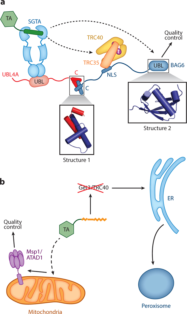
Proper localization of membrane proteins is essential for the function of biological membranes and for the establishment of organelle identity within a cell. Molecular machineries that mediate membrane protein biogenesis need to not only achieve a high degree of efficiency and accuracy, but also prevent off-pathway aggregation events that can be detrimental to cells. The posttranslational targeting of tail-anchored proteins (TAs) provides tractable model systems to probe these fundamental issues. Recent advances in understanding TA-targeting pathways reveal sophisticated molecular machineries that drive and regulate these processes. These findings also suggest how an interconnected network of targeting factors, cochaperones, and quality control machineries together ensures robust membrane protein biogenesis.

Keywords: ATPase; chaperones; membrane protein biogenesis; protein quality control; protein targeting; tail-anchored protein.


Figures

Figure 1
Figure 1
A combination of physicochemical properties in the transmembrane domain (TMD) and C-terminal element (CTE) directs tail-anchored proteins (TAs) to distinct cellular organelles. TAs with weakly hydrophobic TMDs or those with a moderately hydrophobic TMD followed by a basic CTE are targeted to mitochondria or peroxisomes. TAs with moderately to strongly hydrophobic TMDs are targeted to the ER. TAs with long and strongly hydrophobic TMDs further enter vesicular trafficking pathways to reach secretory vesicles, the Golgi apparatus, and the plasma membrane.
Figure 2
Figure 2
Major steps in the yeast GET pathway. (➊) A nascent tail-anchored protein (TA) is captured by Sgt2 after translation by the ribosome. Structure 1 (PDB 3SZ7) shows the Sgt2 tetratricopeptide repeat (TPR) domain, which binds various chaperones. (➋) Sgt2 transfers the TA to Get3, a process stimulated by the Get4/5 complex. Structure 2 (PDB 2LXC) shows the N-terminal domain of Sgt2 bound to the ubiquitin-like (UBL) domain of Get5. (➌) The Get3•TA complex dissociates from Get4/5, and ATP hydrolysis is activated. (➍) The Get2 subunit in the Get1/2 receptor captures the Get3•TA complex. (➎) Following ADP release, Getl interacts with and disassembles the Get3•TA complex, and the TAis inserted into the membrane through an unknown mechanism. (➏) ATP and Get4/5 together drive the release of Get3 from Getl, recycling Get3 for additional rounds of targeting. Structure 3 (PDB 2LNZ) shows the Get5 homodimerization domain. TMD denotes transmembrane domain. Individual proteins are denoted by the following colors: Getl, cyan; Get2, violet; Get3, yellow; Get4, orange; Get5, red; Sgt2, blue; TA, green.
Figure 3
Figure 3
The Get3 ATPase cycle is driven by nucleotides, effectors, and tail-anchored protein (TA) substrates. Structure 1 shows apo-Get3 in an open conformation (PDB 3H84), although whether this species exists in vivo is unclear. (➊) ATP binding induces Get3 into a closed conformation (Structure 2; PDB 2WOJ). (➋) Get4/5 preferentially binds closed Get3 and inhibits its ATPase activity, generating an occluded state (Structure 3; PDB 4PWX). (➌) TA binding induces Get3 into an activated state, and Get3 dissociates from Get4/5. The crystal structure of Get3 bound with a transmembrane domain (TMD) peptide is shown in Structure 4 (PDB 4XTR), although the structural basis for the TA-induced activation of Get3 is unclear. (➍) Activated Get3 hydrolyzes ATP, and the ADP-bound Get3TA complex can bind Get2. (➎) ADP release induces additional rearrangements in the Get3 •TA complex, which enable it to interact with Get1. (➏) The strong preference of Get1 for a wide-open Get3 (Structure 5; PDB 3SJB) drives the release of TA from Get3. CD denotes cytosolic domain. The two subunits in the Get3 homodimer are in blue and tan in all structures.
Figure 4
Figure 4
Two distinct pathways target tail-anchored proteins to peroxisomes. In the direct pathway (left path), Pex19 captures PEX26 in the cytosol and delivers it to peroxisomes via interaction with Pex3. PEX26 is then inserted into the peroxisomal membrane via an unknown mechanism. In the ER-dependent pathway (right path), Pex15 is first targeted to the ER membrane and is then sorted to peroxisomal ER. Exit of Pex15 from the ER occurs via budding of preperoxisomal vesicles that eventually fuse with peroxisomes, and this process requires Pex3 and Pex19. Structure 1 (PDB 3AJB) shows the crystal structure of the Pex3 cytosolic domain bound to the Pex19 N-terminal peptide.
Figure 5
Figure 5
Connection of tail-anchored protein (TA) targeting to quality control pathways. (a) Architecture of the mammalian TA transfer complex, in which SGTA and TRC40 are linked by the heterotrimeric BAG6 complex comprising UBL4A, TRC35, and BAG6. Dashed lines depict the two potential fates of the TA in this complex: transfer to TRC40 for targeting to the ER and transfer to BAG6 for ubiquitylation and degradation. Structure 1 (PDB 4WWR) shows the crystal structure of the complex between the C-terminal helices of UBL4A and BAG6. Structure 2 (PDB 4DWF) shows the crystal structure of the N-terminal UBL domain of BAG6. (b) A local quality control mechanism at mitochondria based on the AAA-ATPase Mspl on the outer mitochondrial membrane, which degrades Pex15 mislocalized to mitochondria when the GET pathway is disabled. Other abbreviations: NLS, nuclear localization sequence; UBL, ubiquitin like.

Similar articles

Cited by

References

    1. Abell BM, Rabu C, Leznicki P, Young JC, High S. 2007. Post-translational integration of tail-anchored proteins is facilitated by defined molecular chaperones. J. Cell Sci. 120:1743–51 - PubMed
    1. Akopian D, Shen K, Zhang X, Shan SO. 2013. Signal recognition particle: an essential protein-targeting machine. Annu. Rev. Biochem. 82:693–721 - PMC - PubMed
    1. Aviram N, Ast T, Costa EA, Arakel EC, Chuartzman SG, et al. 2016. The SND proteins constitute an alternative targeting route to the endoplasmic reticulum. Nature 540:134–38 - PMC - PubMed
    1. Bae W, Lee YJ, Kim DH, Lee J, Kim S, et al. 2008. AKR2A-mediated import of chloroplast outer membrane proteins is essential for chloroplast biogenesis. Nat. Cell Biol. 10:220–27 - PubMed
    1. Beilharz T, Egan B, Silver PA, Hofmann K, Lithgow T. 2003. Bipartite signals mediate subcellular targeting of tail-anchored membrane proteins in Saccharomyces cerevisiae. J. Biol. Chem. 278:8219–23 - PubMed

Publication types

LinkOut - more resources