Review
. 2018 Apr;1860(4):895-908.
doi: 10.1016/j.bbamem.2017.10.004. Epub 2017 Oct 7.

Applications of sequence coevolution in membrane protein biochemistry

Affiliations
Review

Applications of sequence coevolution in membrane protein biochemistry

John M Nicoludis et al. Biochim Biophys Acta Biomembr. 2018 Apr.

Abstract

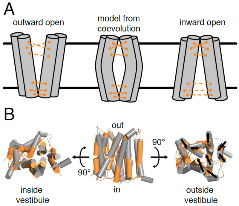
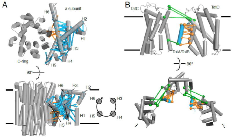
Recently, protein sequence coevolution analysis has matured into a predictive powerhouse for protein structure and function. Direct methods, which use global statistical models of sequence coevolution, have enabled the prediction of membrane and disordered protein structures, protein complex architectures, and the functional effects of mutations in proteins. The field of membrane protein biochemistry and structural biology has embraced these computational techniques, which provide functional and structural information in an otherwise experimentally-challenging field. Here we review recent applications of protein sequence coevolution analysis to membrane protein structure and function and highlight the promising directions and future obstacles in these fields. We provide insights and guidelines for membrane protein biochemists who wish to apply sequence coevolution analysis to a given experimental system.

Keywords: ABC transporters; Clustered protocadherin; Conformational changes; Membrane proteins; Protein-protein interactions; Sequence coevolution analysis.


Figures

Figure 1
Figure 1. Compensatory mutations produce evolutionary couplings in sequence alignments that can identify contacting residue pairs in protein structures
(A) A phenylalanine residue engages in van der Waals interactions with a nearby isoleucine. Random mutation of phenylalanine to glutamine disrupts this interaction, destabilizing the protein. A subsequent mutation of isoleucine to serine restabilizes the interaction through a hydrogen bond (dashed line). (B) Sequence coevolution analysis detects pairs and sets of positions where amino acid identity covaries (e.g. the Phe-Ile to Gln-Ser pairs in orange and cyan, respectively) in a multiple sequence alignment. (C) Coevolving residue pairs can be plotted on a contact map to show overlap of predicted contacts (i.e. coevolving pairs; orange) and true contacts from a high-resolution experimental structure (gray). In α-helical membrane proteins, helical packing results in diagonal signals that can be used to determine which helices contact each other in a parallel (e.g. helices 1 and 3 here) or antiparallel (e.g. helices 1 and 2) interface. (D) Coevolving residue pairs can be used to determine 3-dimensional structures of membrane proteins. Here the coevolving pairs are indicated with dumbbell connections.
Figure 2
Figure 2. Direct methods provide useful structural insights for membrane proteins with diverse folds
(A) The structure of the membrane protein insertase YidC was predicted using plmDCA [41]. This predicted structure positioned the helical paddle domain (HPD; cyan) at the membrane interface. In combination with a concurrently solved crystal structure (illustrated here; PDB ID: 3WO6 [42]), the predicted structure thus suggested dynamics of the HPD (red arrow). (B) EVFold_membrane accurately models membrane helix topology (light blue) compared to the solved structure (gray; PDB ID: 5TCX). Coevolving pairs (orange) confirmed the unusual membrane topology showing close interactions within transmembrane segment pairs S1-S2 and S3-S4 but relatively few contacts between these pairs [43]. (C) EVFold can predict outer membrane β-barrel protein structures, as shown by the agreement between the predicted structure (magenta) and experimental structure (gray; PDB ID: 1P4T) of the Neisseria meningitidis surface protein A, NspA [23]. Note that although the β barrel is accurately predicted, the loop regions are often sparse of coevolving pairs and thus still challenging to predict.
Figure 3
Figure 3. Sequence coevolution can identify functionally important residues
(A) Comparative analysis of dimerizing chemokine GPCRs and non-dimerizing rhodopsin-like GPCRs using SCA reveals a protein sector that is specific to GPCR dimerization (cyan), mapped here on the CXCR4 structure (PDB ID: 3ODU) [66]. (B) mfDCA of BamA identified a coevolving pair (R661-D740) that forms a salt-bridge, as demonstrated in the recent BamA crystal structure (magenta; PDB ID: 4N75). The original structure of the BamA paralog FhaC (gray; PDB ID: 2QDZ), incorrectly modeled the loop containing R661. Reprocessing of the original FhaC dataset with a higher resolution model (cyan; PDB: 4QKY) has the same conformation for this loop as BamA [74,75,77,78]. (C) Differences in helix topology and coevolutionary network between voltage-gated channels and TRP channels, such as the twisting of the S1-S4 domain, the conformational independence of the arginine-bearing S4 of voltage-gated channels, and the allosteric communication between the sensor and pore domains, are supported by coevolving residue pairs [80,83].
Figure 4
Figure 4. Coevolution can predict homomer architecture when the protomer structure is known
(A) Coevolving pairs can represent intramolecular contacts in a monomeric structure or interacting residues across a homomer interface, which complicates structural modeling. (B) The contact map of the first 4 extracellular cadherin (EC) repeat domains of clustered protocadherins shows the intramolecular contacts (gray), dimerization contacts from crystal structures of various isoforms (blue), and coevolving pairs (black) that overlap with either the intramolecular or dimer contacts. (C) Intermolecular coevolving residue pairs (orange) agree with the PcdhγB3 EC1-4 homodimer structure (PDB ID: 5K8R).
Figure 5
Figure 5. Evolutionary couplings of multiple conformations complicates structural modeling of alternating-access transporters
(A) Some coevolving pairs in alternating-access transporters only contact each other in either the outward-open or inward-open conformations. Structural modeling of these transporters using all evolutionary couplings can generate an occluded structure (middle). (B) The outward-open crystal structure of YddG (gray; PDB ID: 5I20) differs from the EVFold_membrane predicted structure (orange) in that the transmembrane segments of the predicted structure are bent inwards (shown with black arrows), closing off the outer vestibule [109].
Figure 6
Figure 6. Coevolving residue pairs help model large membrane protein complexes through an integrative approach
(A) In low resolution EM structures of the ATP synthase, coevolving residue pairs helped determine the registry of the a subunit against the C-ring (orange) and on the clockwise topology of the a subunit helices (cyan) [134,135]. At the top, the structure (the bovine mitochondrial ATP synthase; PDB ID: 5ARA) is viewed from outer side of the membrane, while at the bottom, the structure is view from within the plan of the membrane. (B) Coevolving residue pairs (orange) indicate where TatA or TatB (cyan; PDB ID: 2MN7) dimerizes with TatC (gray; PDB ID: 2HTS). Other coevolving residue pairs (green) suggest oligomerization of the TatA/TatB-TatC complex [145], which, if propagated, may represent formation of the translocation pore, as shown below when the complex is viewed from outside the cell.

Similar articles

Cited by

References

    1. Ivankov DN, Finkelstein AV, Kondrashov Fa. A structural perspective of compensatory evolution. Curr Opin Struct Biol. 2014;26C:104–112. doi: 10.1016/j.sbi.2014.05.004. - DOI - PMC - PubMed
    1. Coucke A, Uguzzoni G, Oteri F, Cocco S, Monasson R, Weigt M. Direct coevolutionary couplings reflect biophysical residue interactions in proteins. J Chem Phys. 2016;145:1–17. doi: 10.1063/1.4966156. - DOI - PubMed
    1. Talavera D, Lovell SC, Whelan S. Covariation is a poor measure of molecular coevolution. Mol Biol Evol. 2015;32:2456–2468. doi: 10.1093/molbev/msv109. - DOI - PMC - PubMed
    1. Göbel U, Sander C, Schneider R, Valencia A. Correlated Mutations and Residue Contacts in Proteins. Proteins Struct Funct Genet. 1994;18:309–317. - PubMed
    1. Korber BTM, Farber M, Robert, Wolpert DH, Lapedes AS. Covariation of mutations in the V3 loop of human immunodeficiency virus type 1 envelope protein : An information theoretic analysis. Proc Natl Acad Sci U S A. 1993;90:7176–7180. - PMC - PubMed

Publication types

Substances

LinkOut - more resources