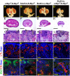
Myc cooperates with β-catenin to drive gene expression in nephron progenitor cells
- PMID: 28993399
- PMCID: PMC5719246
- DOI: 10.1242/dev.153700
Myc cooperates with β-catenin to drive gene expression in nephron progenitor cells
Abstract
For organs to achieve their proper size, the processes of stem cell renewal and differentiation must be tightly regulated. We previously showed that in the developing kidney, Wnt9b regulates distinct β-catenin-dependent transcriptional programs in the renewing and differentiating populations of the nephron progenitor cells. How β-catenin stimulated these two distinct programs was unclear. Here, we show that β-catenin cooperates with the transcription factor Myc to activate the progenitor renewal program. Although in multiple contexts Myc is a target of β-catenin, our characterization of a cell type-specific enhancer for the Wnt9b/β-catenin target gene Fam19a5 shows that Myc and β-catenin cooperate to activate gene expression controlled by this element. This appears to be a more general phenomenon as we find that Myc is required for the expression of every Wnt9b/β-catenin progenitor renewal target assessed as well as for proper nephron endowment in vivo This study suggests that, within the developing kidney, tissue-specific β-catenin activity is regulated by cooperation with cell type-specific transcription factors. This finding not only provides insight into the regulation of β-catenin target genes in the developing kidney, but will also advance our understanding of progenitor cell renewal in other cell types/organ systems in which Myc and β-catenin are co-expressed.
Keywords: Fam19a5; Mouse; Myc; Nephron progenitors; Stem cells; Wnt; β-Catenin.
© 2017. Published by The Company of Biologists Ltd.
Conflict of interest statement
Competing interestsThe authors declare no competing or financial interests.
Figures


Comment in
-
Developmental biology: Renewal of NPCs requires MYC and β-catenin.Nat Rev Nephrol. 2017 Dec;13(12):723. doi: 10.1038/nrneph.2017.151. Epub 2017 Oct 30. Nat Rev Nephrol. 2017. PMID: 29081512 No abstract available.
Similar articles
-
Disparate levels of beta-catenin activity determine nephron progenitor cell fate.Dev Biol. 2018 Aug 1;440(1):13-21. doi: 10.1016/j.ydbio.2018.04.020. Epub 2018 Apr 26. Dev Biol. 2018. PMID: 29705331 Free PMC article.
-
Canonical Wnt9b signaling balances progenitor cell expansion and differentiation during kidney development.Development. 2011 Apr;138(7):1247-57. doi: 10.1242/dev.057646. Epub 2011 Feb 24. Development. 2011. PMID: 21350016 Free PMC article.
-
A β-catenin-driven switch in TCF/LEF transcription factor binding to DNA target sites promotes commitment of mammalian nephron progenitor cells.Elife. 2021 Feb 15;10:e64444. doi: 10.7554/eLife.64444. Elife. 2021. PMID: 33587034 Free PMC article.
-
WNT/beta-catenin signaling in nephron progenitors and their epithelial progeny.Kidney Int. 2008 Oct;74(8):1004-8. doi: 10.1038/ki.2008.322. Epub 2008 Jul 16. Kidney Int. 2008. PMID: 18633347 Free PMC article. Review.
-
Nephron progenitor cell commitment: Striking the right balance.Semin Cell Dev Biol. 2019 Jul;91:94-103. doi: 10.1016/j.semcdb.2018.07.017. Epub 2018 Jul 30. Semin Cell Dev Biol. 2019. PMID: 30030141 Review.
Cited by
-
Identification of differentially expressed genes in cervical cancer by bioinformatics analysis.Oncol Lett. 2018 Aug;16(2):2549-2558. doi: 10.3892/ol.2018.8953. Epub 2018 Jun 12. Oncol Lett. 2018. PMID: 30013649 Free PMC article.
-
Inhibition of GSK3 Represses the Expression of Retinoic Acid Synthetic Enzyme ALDH1A2 via Wnt/β-Catenin Signaling in WiT49 Cells.Front Cell Dev Biol. 2020 Mar 17;8:94. doi: 10.3389/fcell.2020.00094. eCollection 2020. Front Cell Dev Biol. 2020. PMID: 32258025 Free PMC article.
-
Impact of gestational low-protein intake on embryonic kidney microRNA expression and in nephron progenitor cells of the male fetus.PLoS One. 2021 Feb 5;16(2):e0246289. doi: 10.1371/journal.pone.0246289. eCollection 2021. PLoS One. 2021. PMID: 33544723 Free PMC article.
-
The core SWI/SNF catalytic subunit Brg1 regulates nephron progenitor cell proliferation and differentiation.Dev Biol. 2020 Aug 15;464(2):176-187. doi: 10.1016/j.ydbio.2020.05.008. Epub 2020 Jun 3. Dev Biol. 2020. PMID: 32504627 Free PMC article.
-
Can Adipokine FAM19A5 Be a Biomarker of Metabolic Disorders?Diabetes Metab Syndr Obes. 2024 Apr 10;17:1651-1666. doi: 10.2147/DMSO.S460226. eCollection 2024. Diabetes Metab Syndr Obes. 2024. PMID: 38616989 Free PMC article. Review.
References
-
- Bates C. M., Kharzai S., Erwin T., Rossant J. and Parada L. F. (2000). Role of N-myc in the developing mouse kidney. Dev. Biol. 222, 317.- . - PubMed
-
- Boyle S., Misfeldt A., Chandler K. J., Deal K. K., Southard-Smith E. M., Mortlock D. P., Baldwin H. S. and deCaestecker M. (2008). Fate mapping using Cited1-CreERT2 mice demonstrates that the cap mesenchyme contains self-renewing progenitor cells and gives rise exclusively to nephronic epithelia. Dev. Biol. 313, 234-245. 10.1016/j.ydbio.2007.10.014 - DOI - PMC - PubMed
Publication types
MeSH terms
Substances
Grants and funding
LinkOut - more resources
Full Text Sources
Other Literature Sources
Medical
Molecular Biology Databases

