. 2017 Nov 15;130(22):3918-3932.
doi: 10.1242/jcs.207209. Epub 2017 Oct 9.

A functional analysis of TOEFAZ1 uncovers protein domains essential for cytokinesis in Trypanosoma brucei

Affiliations

A functional analysis of TOEFAZ1 uncovers protein domains essential for cytokinesis in Trypanosoma brucei

Amy N Sinclair-Davis et al. J Cell Sci. .

Abstract

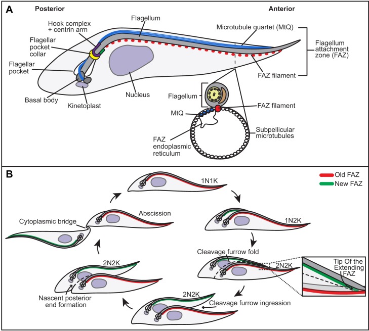
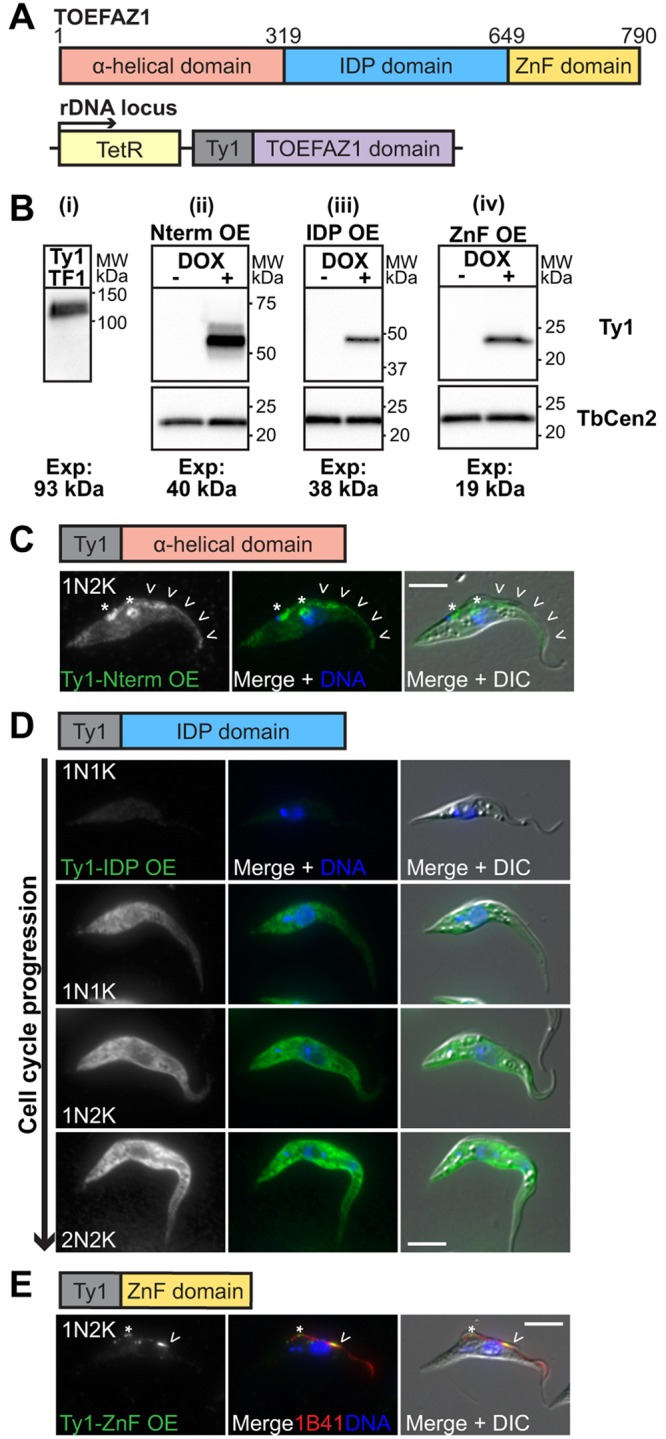
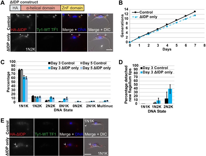
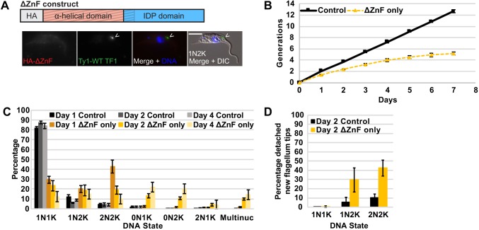
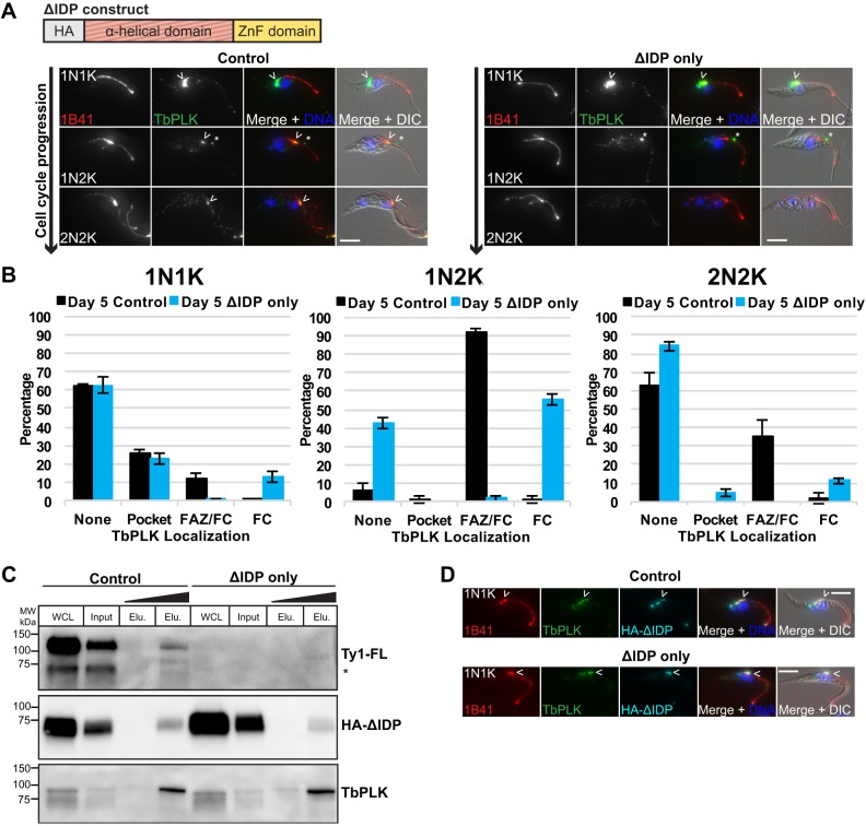
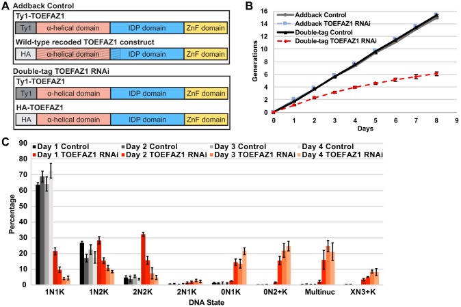
The parasite Trypanosoma brucei is highly polarized, including a flagellum that is attached along the cell surface by the flagellum attachment zone (FAZ). During cell division, the new FAZ positions the cleavage furrow, which ingresses from the anterior tip of the cell towards the posterior. We recently identified TOEFAZ1 (for 'Tip of the Extending FAZ protein 1') as an essential protein in trypanosome cytokinesis. Here, we analyzed the localization and function of TOEFAZ1 domains by performing overexpression and RNAi complementation experiments. TOEFAZ1 comprises three domains with separable functions: an N-terminal α-helical domain that may be involved in FAZ recruitment, a central intrinsically disordered domain that keeps the morphogenic kinase TbPLK at the new FAZ tip, and a C-terminal zinc finger domain necessary for TOEFAZ1 oligomerization. Both the N-terminal and C-terminal domains are essential for TOEFAZ1 function, but TbPLK retention at the FAZ is not necessary for cytokinesis. The feasibility of alternative cytokinetic pathways that do not employ TOEFAZ1 are also assessed. Our results show that TOEFAZ1 is a multimeric scaffold for recruiting proteins that control the timing and location of cleavage furrow ingression.

Keywords: Cleavage furrow ingression; Cytokinesis; Cytoskeleton; Flagellum attachment zone; Polo-like kinase; Trypanosoma brucei.


Conflict of interest statement

Competing interestsThe authors declare no competing or financial interests.

Figures

Fig. 1.
Fig. 1.
Schematic of the cytoskeleton and cell cycle of Trypanosoma brucei. (A) Key cytoskeletal structures involved in this work. (B) The cell cycle in T. brucei. The new flagellum and FAZ are nucleated near the posterior of the cell and are elongated during kinetoplast replication and mitosis. Cleavage furrow ingression initiates at the anterior tip of the extending FAZ, which demarcates the most anterior position of the new-flagellum-containing daughter cell.
Fig. 2.
Fig. 2.
TOEFAZ1 localizes to the tip of the new FAZ and the cleavage furrow. (A) Cells were methanol-fixed and labeled with 1B41 (red), anti-FAZ1 (green), and anti-HA (HA-TOEFAZ1; cyan) antibodies. Arrowheads point to TOEFAZ1 localization. The asterisk in panel ii indicates the new FAZ tip labeled by 1B41. (B) The same cell line as in A was methanol-fixed and stained with 1B41 (red), anti-TbPLK (green) and anti-HA (HA-TOEFAZ1; cyan) antibodies. Asterisks in panels iii–iv point to TbPLK FC labeling; filled-in arrowheads indicate TbPLK FAZ labeling, and empty arrowheads indicate HA-TOEFAZ1 localization. Arrows indicate the colocalization of TbPLK and TOEFAZ1 at the tip of the extending FAZ. DAPI was used to label DNA (blue). Scale bars: 5 µm.
Fig. 3.
Fig. 3.
Overexpression of TOEFAZ1 domains reveals separable functions. (A) Schematic of TOEFAZ1 domain organization (top) and the doxycycline-inducible domain overexpression strategy (bottom). (B) Anti-Ty1 western blots of 427 cells expressing 3×-Ty1–TOEFAZ1 from the endogenous locus (i) and 29-13 cells containing the 3×-Ty1 tagged domain overexpression (OE) constructs that were treated with either 70% ethanol (–DOX) or doxycycline (+DOX) for 1 day (ii–iv). Blots ii–iv were stripped and re-probed with TbCentrin2 (TbCen2) as a loading control. The expected mass of each construct (tag plus domain) is indicated. (C–E) Overexpression cell lines were induced as in B and collected for anti-Ty1 labeling immunofluorescence studies (Ty1-TOEFAZ1 domain OE; green). DAPI was used to label DNA (blue). Asterisks indicate ring-like structures and arrowheads mark FAZ labeling. (C) N-terminus domain cells were fixed with paraformaldehyde. (D) IDP domain cells were fixed with paraformaldehyde. (E) ZnF domain cells were fixed with methanol and stained with 1B41 to mark the FAZ (red). Scale bars: 5 µm.
Fig. 4.
Fig. 4.
The TOEFAZ1 N-terminal domain is required for stable localization to the new FAZ tip. (A) Schematic of TOEFAZ1 RNAi complementation strategy. Gray stripes indicate the recoded segment of the gene. (B) ΔN-terminus cells in the endogenously tagged 3X-Ty1 TOEFAZ1 RNAi background were treated with vehicle control or doxycycline (ΔN-terminus only) for 1 day. Cells were methanol-fixed and labeled with anti-HA (HA-ΔN-terminus; red), anti-Ty1 [Ty1-WT TOEFAZ1 (TF1); green] and DAPI for DNA (blue). Arrowheads indicate TOEFAZ1 construct localization. (C) The ΔN-terminus cell line was treated as in B and cell number was monitored every 24 h. The curve represents the mean of three independent experiments. (D) Control and ΔN-terminus only cells from C were paraformaldehyde-fixed and stained with DAPI for DNA (blue) after 2 days of TOEFAZ1 depletion. Asterisks indicate a detached new flagellum tip. (E) Quantification of DNA state in control cells (black and gray) and ΔN-terminus only cells (dark and light red). 300 cells were counted per condition for three independent experiments. (F) Quantification of detached new flagellum tips after 2 days of TOEFAZ1 depletion in control and ΔN-terminus only cells as in E. Scale bars: 5 µm. Error bars are the s.d.
Fig. 5.
Fig. 5.
The ΔIDP construct complements WT TOEFAZ1 function. The same analyses as in Fig. 4 were performed on the ΔIDP cell line. WT TF1, WT TOEFAZ1. (A) ΔIDP cells were fixed after 2 days of TOEFAZ1 RNAi and labeled as in Fig. 4B. The asterisk indicates posterior labeling by the ΔIDP protein. (B) ΔIDP cells were induced and cell growth was monitored. The curve is the mean of three independent experiments. (C) Quantification of DNA state after TOEFAZ1 depletion in control (black and gray) and ΔIDP only (dark and light blue) cells. 300 cells were counted for each condition for three independent experiments. (D) Quantification of detached new flagellum tips in control and ΔIDP-only cells after 3 days of TOEFAZ1 depletion as in C. (E) Control and ΔIDP-only cells after 2 days of TOEFAZ1 depletion were methanol-fixed and labeled as in A. Arrowheads in A and E indicate ΔIDP protein labeling in the cell posterior. Scale bars: 5 µm. Error bars are the s.d.
Fig. 6.
Fig. 6.
The ΔZnF construct does not localize to the new FAZ tip. The ΔZnF cell line was analyzed as in Fig. 4. WT TF1, WT TOEFAZ1. (A) Uninduced ΔZnF cells were methanol-fixed and prepared as in Fig. 4B. The arrowhead indicates WT TOEFAZ1 localization. (B) TOEFAZ1 RNAi was induced in ΔZnF cells and cell growth was monitored. The curve is the mean of three independent experiments. (C) Quantification of DNA state in control (black and gray) and ΔZnF-only cells (gold and yellow) after WT TOEFAZ1 depletion. 300 cells were counted per condition for three independent experiments. (D) The percentage of detached new flagellum tips was quantified in control and ΔZnF-only cells after 2 days of TOEFAZ1 depletion as in C. Scale bar: 5 µm. Error bars are the s.d.
Fig. 7.
Fig. 7.
The IDP domain is required to maintain TbPLK localization at the new FAZ tip. (A) Control and ΔIDP-only cells were methanol-fixed and labeled with 1B41 (red) and anti-TbPLK (green) after 5 days of TOEFAZ1 depletion. Arrowheads indicate TbPLK localization. Asterisks mark TbPLK FC labeling. (B) Quantification of TbPLK localization in cells from A. 300 cells per condition from three independent experiments were counted. Error bars are the s.d. (C) TOEFAZ1 RNAi was induced in ΔIDP cells for 3 days and cells were collected for TbPLK immunoprecipitation. Both input and immunoprecipitated fractions were western blotted with antibodies against Ty1 (Ty1-FL TOEFAZ1), HA (HA-ΔIDP) and TbPLK. Elution fractions are equal volume (1.2%) and 20% elution, respectively. Asterisk indicates degradation product. WCL, whole-cell lysate. (D) ΔIDP cells were methanol-fixed and stained with 1B41 (red), anti-TbPLK (green), and anti-HA (HA-ΔIDP; cyan) after 2 days of WT TOEFAZ1 depletion. Arrowheads indicate ΔIDP and TbPLK colocalization at the short new FAZ tip. DAPI was used to stain DNA (blue). Scale bars: 5 µm.
Fig. 8.
Fig. 8.
TOEFAZ1 RNAi causes severe cytokinetic defects. All analyses are the same as in Fig. 4. (A) Schematic of the TOEFAZ1 endogenous locus in addback control (top) and double-tagged TOEFAZ1 RNAi cell lines (bottom). (B) TOEFAZ1 RNAi was induced in the addback control and double-tag RNAi lines, and cell growth was monitored. The curve is the average of three independent experiments. (C) Quantification of DNA state in control (black and gray) and TOEFAZ1-depleted cells (dark and light red) in the double-tag cell line. 300 cells per condition were quantified for three independent experiments. 0N2+K: no nuclei and 2+ kinetoplasts; XN3+K: 1 or 2 nuclei and 3+ kinetoplasts. Error bars are the s.d.

Similar articles

Cited by

References

    1. Al-Zain A., Schroeder L., Sheglov A. and Ikui A. E. (2015). Cdc6 degradation requires phosphodegron created by GSK-3 and Cdk1 for SCFCdc4 recognition in Saccharomyces cerevisiae. Mol. Biol. Cell 26, 2609-2619. 10.1091/mbc.E14-07-1213 - DOI - PMC - PubMed
    1. Anderson W. A. and Ellis R.A. (1965). Ultrastructure of Trypanosoma lewisi: Flagellum, Microtubules, and the Kinetoplast. J. Protozool. Res. 12, 483-499. 10.1111/j.1550-7408.1965.tb03247.x - DOI
    1. Aslett M., Aurrecoechea C., Berriman M., Brestelli J., Brunk B. P., Carrington M., Depledge D. P., Fischer S., Gajria B., Gao X. et al. (2009). TriTrypDB: a functional genomic resource for the Trypanosomatidae. Nucleic Acids Res. 38, D457-D462. 10.1093/nar/gkp851 - DOI - PMC - PubMed
    1. Bah A., Vernon R. M., Siddiqui Z., Krzeminski M., Muhandiram R., Zhao C., Sonenberg N., Kay L. E. and Forman-Kay J. D. (2015). Folding of an intrinsically disordered protein by phosphorylation as a regulatory switch. Nature 519, 106-109. 10.1038/nature13999 - DOI - PubMed
    1. Bajar B. T., Wang E. S., Lam A. J., Kim B. B., Jacobs C. L., Howe E. S., Davidson M. W., Lin M. Z. and Chu J. (2016). Improving brightness and photostability of green and red fluorescent proteins for live cell imaging and FRET reporting. Sci. Rep. 6, 1-12. 10.1038/srep20889 - DOI - PMC - PubMed

MeSH terms

Substances