

Triple-phase helical computed tomography of an arterio-hepatic venous shunt in a hepatic tumor in a dog
- PMID: 28993602
- PMCID: PMC5745169
- DOI: 10.1292/jvms.17-0373
Triple-phase helical computed tomography of an arterio-hepatic venous shunt in a hepatic tumor in a dog
Abstract
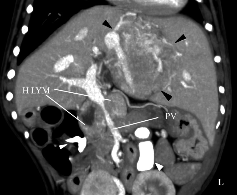
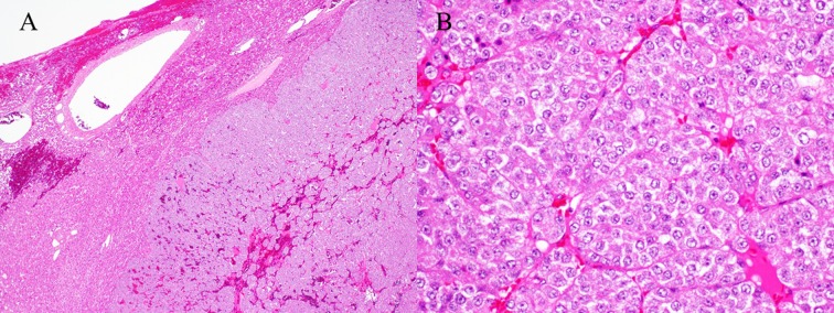
A 10-year-old French bulldog presented with an abdominal tumor. Triple-phase helical computed tomography was performed, revealing a hepatic tumor, an enlarged hepatic lymph node, and no masses in other organs. The hepatic tumor demonstrated marked enhancement, similar to that of the aorta in the arterial phase. The tumor had rich vascularization and a hepatic arterio-venous shunt formed between the hepatic artery and middle hepatic vein. The hepatic tumor was surgically removed and histological diagnosis revealed a hepatic carcinoid tumor. During surgery, rapid massive arterial hemorrhage occurred from the site of the incision. The animal died without improvement post-surgery. In the case of an arterio-venous shunt in a hepatic tumor, it is important to be careful to avoid perioperative bleeding.
Keywords: abdomen; arterio-venous shunt; canine; computed tomography; liver tumor.
Figures


Similar articles
-
Extensive portal-hepatic venous shunts accompanied by arterio-portal shunts.Gastrointest Radiol. 1988 Oct;13(4):351-4. doi: 10.1007/BF01889097. Gastrointest Radiol. 1988. PMID: 3169483
-
High-Flow Arterio-Hepatic Venous Shunt in Hepatocellular Carcinoma: Use of Multi-Electrode Radiofrequency for Shunt Obliteration.Cardiovasc Intervent Radiol. 2015 Oct;38(5):1330-4. doi: 10.1007/s00270-014-0990-2. Epub 2014 Sep 5. Cardiovasc Intervent Radiol. 2015. PMID: 25189667
-
IMAGING DIAGNOSIS-ECTOPIC SPLEEN MIMICKING HEPATIC TUMOR WITH INTRA-ABDOMINAL METASTASES INVESTIGATED VIA TRIPLE-PHASE HELICAL COMPUTED TOMOGRAPHY IN A DOG.Vet Radiol Ultrasound. 2017 May;58(3):E26-E30. doi: 10.1111/vru.12395. Epub 2016 Jul 5. Vet Radiol Ultrasound. 2017. PMID: 27377289
-
Arterio-Hepatic Venous Fistula Following Liver Biopsy: A Rare Case Report and Literature Review.Vasc Endovascular Surg. 2021 Feb;55(2):177-182. doi: 10.1177/1538574420954300. Epub 2020 Sep 3. Vasc Endovascular Surg. 2021. PMID: 32878580 Review.
-
Congenital hepatic shunts.Radiographics. 2004 May-Jun;24(3):755-72. doi: 10.1148/rg.243035046. Radiographics. 2004. PMID: 15143226 Review.
Cited by
-
A case of primary pulmonary paraganglioma in a dog.Open Vet J. 2024 Oct;14(10):2714-2720. doi: 10.5455/OVJ.2024.v14.i10.22. Epub 2024 Oct 31. Open Vet J. 2024. PMID: 39545184 Free PMC article.
References
-
- Breznock E. M., Berger B., Pendray D., Wagner S., Manley P., Whiting P., Hornof W., West D.1983. Surgical manipulation of intrahepatic portocaval shunts in dogs. J. Am. Vet. Med. Assoc. 182: 798–805. - PubMed
-
- Kim H. C., Suk K. T., Kim D. J., Yoon J. H., Kim Y. S., Baik G. H., Kim J. B., Kim C. H., Sung H., Choi J. Y., Han K. H., Park S. H.2014. Transarterial chemoembolization in Barcelona Clinic Liver Cancer Stage 0/A hepatocellular carcinoma. World J. Gastroenterol. 20: 745–754. doi: 10.3748/wjg.v20.i3.745 - DOI - PMC - PubMed
Publication types
MeSH terms
LinkOut - more resources
Full Text Sources
Other Literature Sources
Medical