

Triple-phase helical computed tomography of an arterio-hepatic venous shunt in a hepatic tumor in a dog
- PMID: 28993602
- PMCID: PMC5745169
- DOI: 10.1292/jvms.17-0373
Triple-phase helical computed tomography of an arterio-hepatic venous shunt in a hepatic tumor in a dog
Abstract
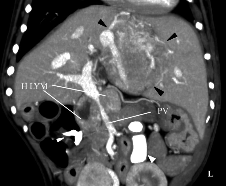
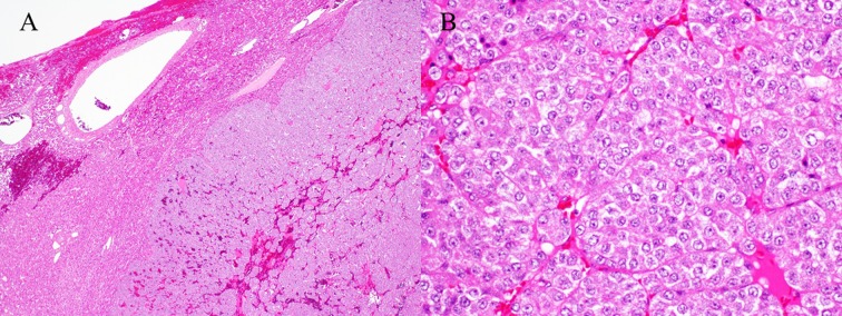
A 10-year-old French bulldog presented with an abdominal tumor. Triple-phase helical computed tomography was performed, revealing a hepatic tumor, an enlarged hepatic lymph node, and no masses in other organs. The hepatic tumor demonstrated marked enhancement, similar to that of the aorta in the arterial phase. The tumor had rich vascularization and a hepatic arterio-venous shunt formed between the hepatic artery and middle hepatic vein. The hepatic tumor was surgically removed and histological diagnosis revealed a hepatic carcinoid tumor. During surgery, rapid massive arterial hemorrhage occurred from the site of the incision. The animal died without improvement post-surgery. In the case of an arterio-venous shunt in a hepatic tumor, it is important to be careful to avoid perioperative bleeding.
Keywords: abdomen; arterio-venous shunt; canine; computed tomography; liver tumor.
Figures


References
-
- Breznock E. M., Berger B., Pendray D., Wagner S., Manley P., Whiting P., Hornof W., West D.1983. Surgical manipulation of intrahepatic portocaval shunts in dogs. J. Am. Vet. Med. Assoc. 182: 798–805. - PubMed
-
- Kim H. C., Suk K. T., Kim D. J., Yoon J. H., Kim Y. S., Baik G. H., Kim J. B., Kim C. H., Sung H., Choi J. Y., Han K. H., Park S. H.2014. Transarterial chemoembolization in Barcelona Clinic Liver Cancer Stage 0/A hepatocellular carcinoma. World J. Gastroenterol. 20: 745–754. doi: 10.3748/wjg.v20.i3.745 - DOI - PMC - PubMed
Publication types
MeSH terms
LinkOut - more resources
Full Text Sources
Other Literature Sources
Medical