. 2017 Dec;2(12):1592-1599.
doi: 10.1038/s41564-017-0033-2. Epub 2017 Oct 9.

Evolution of host adaptation in the Salmonella typhoid toxin

Affiliations

Evolution of host adaptation in the Salmonella typhoid toxin

Xiang Gao et al. Nat Microbiol. 2017 Dec.

Erratum in

Abstract

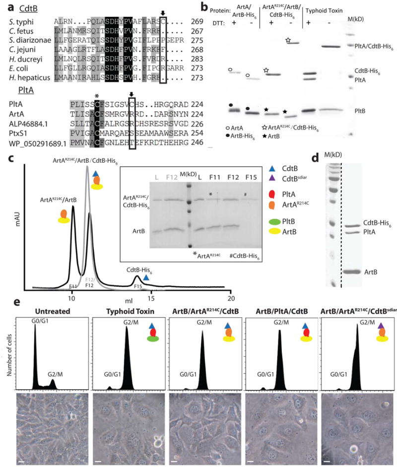
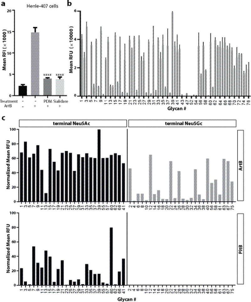
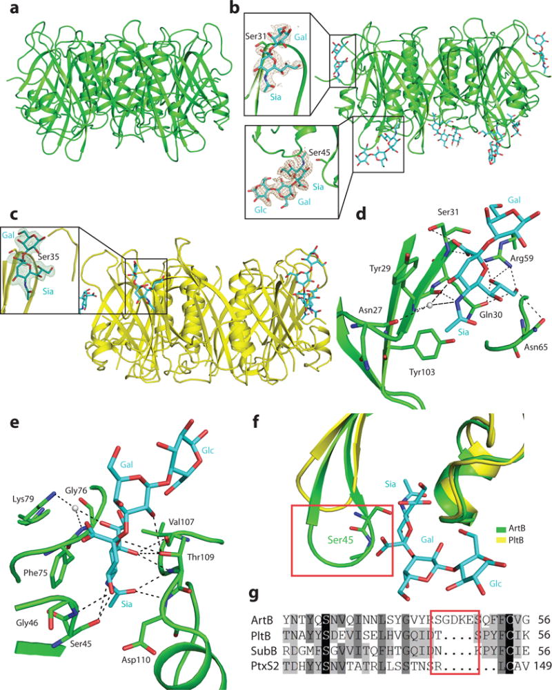
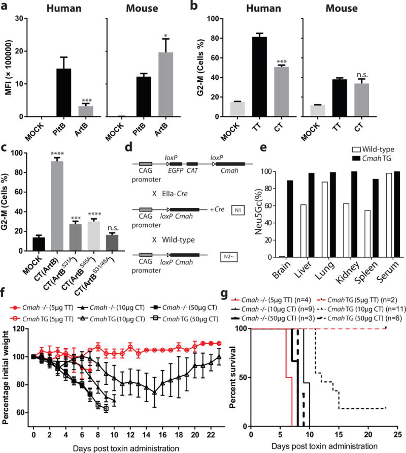
The evolution of virulence traits is central for the emergence or re-emergence of microbial pathogens and for their adaptation to a specific host 1-5 . Typhoid toxin is an essential virulence factor of the human-adapted bacterial pathogen Salmonella Typhi 6,7 , the cause of typhoid fever in humans 8-12 . Typhoid toxin has a unique A2B5 architecture with two covalently linked enzymatic 'A' subunits, PltA and CdtB, associated with a homopentameric 'B' subunit made up of PltB, which has binding specificity for the N-acetylneuraminic acid (Neu5Ac) sialoglycans 6,13 prominently present in humans 14 . Here, we examine the functional and structural relationship between typhoid toxin and ArtAB, an evolutionarily related AB5 toxin encoded by the broad-host Salmonella Typhimurium 15 . We find that ArtA and ArtB, homologues of PltA and PltB, can form a functional complex with the typhoid toxin CdtB subunit after substitution of a single amino acid in ArtA, while ArtB can form a functional complex with wild-type PltA and CdtB. We also found that, after addition of a single-terminal Cys residue, a CdtB homologue from cytolethal distending toxin can form a functional complex with ArtA and ArtB. In line with the broad host specificity of S. Typhimurium, we found that ArtB binds human glycans, terminated in N-acetylneuraminic acid, as well as glycans terminated in N-glycolylneuraminic acid (Neu5Gc), which are expressed in most other mammals 14 . The atomic structure of ArtB bound to its receptor shows the presence of an additional glycan-binding site, which broadens its binding specificity. Despite equivalent toxicity in vitro, we found that the ArtB/PltA/CdtB chimaeric toxin exhibits reduced lethality in an animal model, indicating that the host specialization of typhoid toxin has optimized its targeting mechanisms to the human host. This is a remarkable example of a toxin evolving to broaden its enzymatic activities and adapt to a specific host.


Conflict of interest statement

Competing interests

The authors declare no competing interests

Figures

Figure 1
Figure 1
The ArtAB toxin components can form a functional complex with typhoid toxin subunits. a, Amino acid sequence comparison of CdtB and PltA homologs. Conserved and unique cysteines are indicated with an asterisk and an arrow, respectively. ALP46884.1 and WP_050291689.1 are PltA homologs from Escherichia coli and Yersinia kristensenii, respectively. PltxS1 is the A subunit from pertussis toxin. b, Purified ArtB/ArtA, ArtB/ArtAR214C/CdtB, or typhoid toxin (PltB/PltA/CdtB) protein complexes were analyzed by SDS-PAGE in the presence or absence of DTT to release CdtB, which is linked to the complex by a disulfide bond. The migration of ArtA and His6-tagged CdtB are very similar in SDS-PAGE so in the presence of DTT the two bands overlap. In the absence of DTT, ArtA and CdtB (like CdtB and PltA in the case of typhoid toxin, see right lanes) migrate as a single, slower moving band, an indication that these subunits are linked by a disulfide bond. ○: indicates ArtA; ●: indicates ArtB-His6; ☆: indicates ArtAR214C/CdtB-His6; ★: indicates ArtB. This experiment was carried out twice with equivalent results. c, The ArtAR214C/ArtB/CdtB chimeric toxin complex was analyzed by ion exchange chromatography before (gray) and after (black) treatment with DTT. L: loading control; M: molecular weight markers; F: chromatographic fraction. Inset shows SDS-PAGE analyzes of the indicated chromatographic fractions. *: ArtAR214C; #: CdtB-His6. This experiment was carried out once. d, ArtB can form a complex with wild-type PltA and CdtB. The ArtB/PltA/CdtB complex was purified by ion exchange and size exclusion chromatography and subsequently analyzed by SDS-PAGE and coomassie blue staining. This experiment was carried out three times with equivalent results. The dashed black line indicates that this panel is a composite image of two discontinuous lanes from the same gel. e, Toxicity of the chimeric toxin complexes. Cultured Henle-407 epithelial cells were treated with Typhoid Toxin (3.5 pM), ArtB/ArtAR214A/CdtB (15 pM), ArtB/PltA/CdtB (15 pM), or ArtB/ArtAR214A/CdtBsdiar, and the CdtB-mediated cell cycle arrest was assayed by flow cytometric analysis. CdtBsdiar: CdtB from S. diarizonae. Light microscopic images of mock or toxin treated cells are also shown. Scale bar: 50μm. This experiment was carried out three times with equivalent results.
Figure. 2
Figure. 2
ArtB binds Neu5Ac- and Neu5Gc-terminated glycans. a, Removal of surface glycans reduces ArtB binding to cultured cells. Henle-407 cells were treated with a mixture of glycosidases (PDM, protein deglycosylation mix) or a sialidase (α2–3/6/8-neuraminidase) and the ability of treated and control cells to bind fluorescently labeled ArtB (2.5 μM) was evaluated by flow cytometry. The y-axis values represent the relative fluorescence intensity (RFI). Bars represent mean ± standard deviation of at least three independent measurements. Two-tailed Student’s t tests were performed to determine the statistical significance for two group comparisons. ****: P<0.0001, compare to the relative fluorescence intensity of ArtB-binding to the untreated cells. b, ArtB-binding to a customized glycan microarray. The y-axis values represent average and standard deviation of the relative fluorescence units (RFU) from four independent experiments, and the x-axis indicates the glycan numbers (see also Table S1). c, Comparison of ArtB-binding to paired Neu5Ac- and Neu5Gc- terminated glycans. The y-axis values represent the normalized average relative fluorescence units (RFU) from four independent experiments and the x-axis depicts the glycan numbers (see also Table S2).
Figure. 3
Figure. 3
The atomic structure of ArtB bound to its receptor shows the presence of an additional glycan-binding site. a, Atomic structure of the ArtB pentamer shown as a ribbon cartoon. b, Atomic structure of the ArtB pentamer in complex with the Neu5Acα2–3Galβ1–4Glc oligosaccharide shown as a ribbon cartoon. Cyan, blue and red sticks represent carbon, nitrogen and oxygen atoms in the sugar backbone. Insets show the close-up views of Neu5Acα2–3Galβ1–4Glc and Neu5Acα2–3Gal. Brown mesh represents the sugar composite annealed omit difference density map contoured at 2.0σ. c, Atomic structure of the PltB pentamer in complex with the Neu5Acα2–3Gal oligosaccharide is shown as a ribbon cartoon. Cyan, blue and red sticks represent carbon, nitrogen and oxygen atoms in the sugar backbone. Insets show the close-up views of Neu5Acα2–3Galβ1–4Glc. Green mesh represents the sugar composite annealed omit difference density map contoured at 2.5σ. d and e, Interactions between ArtBSer31 (d) and ArtBSer45 (e) with Neu5Acα2–3Gal and Neu5Acα2–3Galβ1–4Glc, respectively. ArtB is shown as a green colored ribbon cartoon, the sugar and the amino acids interacting with the sugar are shown as sticks, the interactions are shown in black dashes and water is shown as gray balls. f, Structural comparison of ArtBSer45 sugar-binding site with the equivalent surface in PltB. Blue and red sticks in the sugar backbone represent nitrogen and oxygen atoms, respectively. g, Amino acid sequence alignment of ArtBSer45 glycan-binding site with the equivalent regions in PltB, SubB and PtxS2. The red boxes depicted in f and g highlight the insert sequence and the associated structural features that are uniquely present in ArtB.
Figure 4
Figure 4
The ArtB/PltA/CdtB chimeric toxin exhibits reduced lethality in mice relative to typhoid toxin. a, ArtB and PltB (2. 5 μM each) binding to human (Henle-407) or mouse (embryo fibroblasts) cells. The binding of fluorescently-labeled PltB and ArtB was evaluated by flow cytometry. The y-axis values represent the mean fluorescence index (MFI) and are the mean ± SD of at least three independent measurements. *: p < 0.05; ***: p < 0.0005. b, Relative toxicity to human (Henle-407) or mouse (embryo fibroblasts) cells of purified typhoid toxin (TT) (3 pM) and ArtB/PltA/CdtB chimeric toxin (CT) (3 pM). The percentage of cells in G2/M, a measured of CdtB toxic activity, after application of the indicated toxins was determined by flow cytometry. Data are the mean ± SD of at least three independent determinations. ***: p < 0.0005, n.s. (no significance): p>0.05 compared to untreated cells. c, Relative contribution of the different ArtB glycan-biding sites to toxicity. Different ArtB/PltA/CdtB chimeric toxin (CT) preparations containing the indicated ArtB mutants were tested for their ability to intoxicate cultured cells. Equal amounts (250 pM) of chimeric toxin preparations were applied to the cultured cells and the percentage of cells in G2/M was determined by flow cytometry. Data are the mean ± SD of at least three independent determinations. ****: p < 0.0001, ***: p < 0.0005, n.s. (no significance): p>0.05 compared to untreated cells. In a-c two-tailed Student’s t tests were performed to determine the statistical significance for two group comparisons. d, Scheme of the strategy used to generate a Cmah TG mouse. Targeted Cre-inducible Cmah TG mice (transgene insertion in H11 locus, top) were crossed with EIIa-Cre mice that induce Cre expression at the preimplantation embryo stage and the resulting N1 generation mouse with the Cmah transgene and Cre was further mated with wild-type mice. Among the resulting N2 generation, the mice that had Cmah transgene but lacked Cre were selected as systemic Cmah TG mice and maintained by crossing with wild-type mice. e, Tissue homogenates obtained from 11-week-old male mice were hydrolyzed in 2M acetic acid to release sialic acids after treatment with 0.1M sodium hydroxide to remove O-acetylation of sialic acids. The percentage of Neu5Gc in total sialic acids was determined by HPLC using DMB-derivatization method. Each bar represents the average of samples from two mice per genotype. Cmah TG mice showed remarkably high Neu5Gc expression in all tissues tested. f and g, Mouse toxicity of the ArtB/PltA/CdtB chimeric toxin relative to typhoid toxin. Cmah −/− or Cmah transgenic (TG) mice were administered intraorbitally either typhoid toxin (TT) (5μg) or ArtB/PltA/CdtB chimeric toxin (CT) (10 or 50 μg) and their body weight (f) and survival (g) at the indicated times were recorded. Values in (f) are the mean and standard deviations. The difference in weight loss between groups (f) was analyzed by the Mann-Whitney test (TT vs CT in Cmah TG: p <0.0001, TT vs CT in Cmah −/−: p<0.05). The difference in survival (g) was analyzed by the Mantel Cox test. (TT vs CT in Cmah TG: p <0.05; TT (5 μg) vs CT (10 μg) in Cmah −/−: p <0.001.

References

    1. Jackson R, Johnson L, Clarke S, Arnold D. Bacterial pathogen evolution: breaking news. Trends Genet. 2011;27:32–40. - PubMed
    1. Donnenberg MS, Whittam TS. Pathogenesis and evolution of virulence in enteropathogenic and enterohemorrhagic Escherichia coli. J Clin Invest. 2001;107:539–548. - PMC - PubMed
    1. Bentley S, Parkhill J. Genomic perspectives on the evolution and spread of bacterial pathogens. Proc Biol Sci. 2015;282:20150488. - PMC - PubMed
    1. Alizon S, Michalakis Y. Adaptive virulence evolution: the good old fitness-based approach. Trends Ecol Evol. 2015;30:248–254. - PubMed
    1. Daugherty M, Malik H. Rules of engagement: molecular insights from host-virus arms races. Annu Rev Genet. 2012;46:677–700. - PubMed

MeSH terms