. 2017 Oct 9;7(1):12807.
doi: 10.1038/s41598-017-12718-4.

Dissecting the mycobacterial cell envelope and defining the composition of the native mycomembrane

Affiliations

Dissecting the mycobacterial cell envelope and defining the composition of the native mycomembrane

Laura Chiaradia et al. Sci Rep. .

Abstract

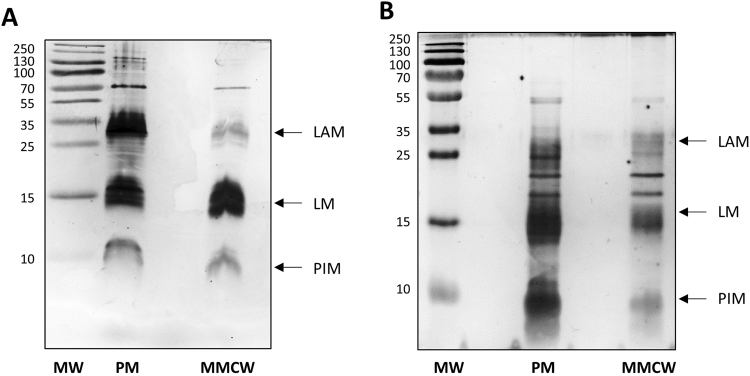
The mycobacterial envelope is unique, containing the so-called mycomembrane (MM) composed of very-long chain fatty acids, mycolic acids (MA). Presently, the molecular composition of the MM remains unproven, due to the diversity of methods used for determining its composition. The plasma membranes (PM) and the native MM-containing cell walls (MMCW) of two rapid-growing mycobacterial species, Mycobacterium aurum and M. smegmatis, were isolated from their cell lysates by differential ultracentrifugation. Transmission electron microscopy and biochemical analyses demonstrated that the two membranes were virtually pure. Bottom-up quantitative proteomics study indicated a different distribution of more than 2,100 proteins between the PM and MMCW. Among these, the mannosyltransferase PimB, galactofuranosyltransferase GlfT2, Cytochrome p450 and ABC transporter YjfF, were most abundant in the PM, which also contain lipoglycans, phospholipids, including phosphatidylinositol mannosides, and only a tiny amount of other glycolipids. Antigen85 complex proteins, porins and the putative transporters MCE protein family were mostly found in MMCW fraction that contains MA esterifying arabinogalactan, constituting the inner leaflet of MM. Glycolipids, phospholipids and lipoglycans, together with proteins, presumably composed the outer leaflet of the MM, a lipid composition that differs from that deduced from the widely used extraction method of mycobacterial cells with dioctylsulfosuccinate sodium.


Conflict of interest statement

The authors declare that they have no competing interests.

Figures

Figure 1
Figure 1
A tentative model of the arrangement of the cell envelope of M. smegmatis. (adapted from). The outermost layer (OL) is primarily constituted of proteins, with small amounts of carbohydrate and only a tiny amount of lipids. The cell wall is a giant tripartite complex composed of the outer membrane, the so-called mycomembrane (MM), arabinogalactan (AG) and peptidoglycan (PG). The inner leaflet of the MM is made of very long-chain fatty acids (mycolic acids) esterifying AG, which in turn is covalently attached to PG. The outer leaflet of the MM is presumably composed of lipids extractable with organic solvents, which include phospholipids, trehalose mycolates, glycopeptidolipids, and lipoglycans. A periplasmic space separates the cell wall from the conventional lipid bilayer PM of phospholipids and proteins whose thickness is surprisingly similar to that of MM, around 7–8 nm, despite the presence of the very long chain mycolic acids. The scales of the various cell envelope compartments are based on the data from CEMOVIS, except that of the OL, which is adapted from cryo-microscopy. For clarity, molecules are not drawn on scale. TMM : trehalose monomycolates; TDM: trehalose dimycolates; GPL: glycopeptidolipids; PL: phospholipids; PIM: phosphatidyl-myo-inositol-mannosides ; LAM: lipoarabinomannans; TAG: triacylglycerols; Ag85: antigen 85.
Figure 2
Figure 2
A schematic representation of the fractionation of the mycobacterial lysate for isolating the MMCW and the PM. After mechanical breaking, the bacterial lysate was centrifuged to yield crude cell wall fraction (pellet P10), which was then layered on a sucrose step gradient and centrifuged to recover the cell walls. The S10 supernatant was centrifuged and the S27 supernatant was used to collect the crude PM fraction (pellet P100), which was then layered on a sucrose step gradient.
Figure 3
Figure 3
Isolation and NADH oxidase activity of mycobacterial membranes. Visualization of the F1 and F2 of Mau (A) and Msm (D). Negative staining of the F1 and F2 fractions of Mau (B) and Msm (E); bars represent 200 nm for F1 and 1 µm for F2. NADH oxidase activity of F1 (black symbols) and F2 (grey symbols) of Mau (C) and Msm (F).
Figure 4
Figure 4
Biochemical characterization of mycobacterial membrane markers. Purified F1 and F2 of Mau (A) and Msm (B) were hydrolyzed, derivatized and analyzed by gas chromatography-mass spectrometry, and their contents (nmoles per mg of dried membranes) in galactose (Gal), arabinose (Ara), mannose (Man), glucosamine (Gln), muramic acid (Mur. Ac.) and diaminopimelic acid (DAP) were determined. F1 (grey symbols) and F2 (black symbols).
Figure 5
Figure 5
Analysis of mycobacterial membrane proteins. SDS-PAGE analysis of proteins extracted from the F1 (PM) and F2 (mycomembrane-containing cell walls, MMCW) of Mau (A) and Msm (B). Western blotting of PM and MMCW fractions from Mau (upper panel) and Msm (lower panel) using anti-Antigen85 (C) and anti-ATP synthase beta, AtpD (D) antibodies. Western blotting of PM and MMCW fractions from Msm using anti-MspA antibodies (E). MW: molecular weight markers in kDa.
Figure 6
Figure 6
Quantitative proteomics analysis of M. smegmatis proteins. Volcano plot presentation of the statistical significance of Msm protein abundances as a function of protein abundance ratios between PM and MMCW fractions. Horizontal lines depict a p < 0.01 cutoff and vertical lines depict 1/3- and 3-fold ratios cutoffs. Each dot corresponds to a single identified protein. Dark grey dots indicate proteins enriched in each fraction. Proteins considered as markers of each membrane fraction and significantly enriched are represented in the volcano plot with red triangles. The corresponding protein name and gene number in parentheses are indicated.
Figure 7
Figure 7
Analysis of mycobacterial membrane lipids. High performance thin-layer chromatography of lipids extracted from the PM and MMCW of Mau (A) and Msm (B). The plates were developed in CHCl3/CH3OH/H2O (65:25:4, v/v/v) and lipid spots were revealed by immerging the plate in primuline. In C: a TLC of lipids extracted from PM, MMCW and bacteria (TEL, total extractable lipids) of Msm. The plate was developed in CHCl3/CH3OH (9:1, v/v), and revealed with anthrone, followed by heating. The GPL are colored in blue on the plate. (D) Analysis of TAG: TLC were developed in Petroleum Ether /Diethyl Ether (9:1, v/v) and revealed by immerging the plate in a primuline bath. 100 µg of lipid mixtures were deposited on HPTLC plates. AL: apolar lipids; CL: cardiolopin; PG: phosphatidyl glycerol; PE: phosphatidyl ethanolamine; PI: phosphatidyl inositol; PIM: phosphatidyl-myo-inositol mannosides; TMM: trehalose monomycolates; TDM: trehalose dimycolates; GMM: glucose monomycolates; GPL: glycopeptidolipids; PL: phospholipids; TAG: triacylglycerols.
Figure 8
Figure 8
Analysis of mycobacterial membrane lipoglycans. SDS-PAGE analysis of lipoglycans from the PM and MMCW of Mau (A) and Msm (B). F1 and F2 fractions were digested with proteases prior to their analysis. LAM: lipoarabinomannan; LM: lipomannan; PIM: phosphatidyl-myo-inositol mannosides. MW: molecular weight markers in kDa.

References

    1. Han XY, et al. A new Mycobacterium species causing diffuse lepromatous leprosy. Am J Clin Pathol. 2008;130:856–864. doi: 10.1309/AJCPP72FJZZRRVMM. - DOI - PubMed
    1. Wayne LG, Kubica GP. The Mycobacteria. The Bergey’s manual of systematic bacteriology. 1986;2:1435–1457.
    1. Daffe M, Draper P. The envelope layers of mycobacteria with reference to their pathogenicity. Adv Microb Physiol. 1998;39:131–203. doi: 10.1016/S0065-2911(08)60016-8. - DOI - PubMed
    1. Brennan PJ, Goren MB. Structural studies on the type-specific antigens and lipids of the mycobacterium avium. Mycobacterium intracellulare. Mycobacterium scrofulaceum serocomplex. Mycobacterium intracellulare serotype 9. J Biol Chem. 1979;254:4205–4211. - PubMed
    1. Jarlier V, Nikaido H. Permeability Barrier to Hydrophilic Solutes in Mycobacterium-Chelonei. J Bacteriol. 1990;172:1418–1423. doi: 10.1128/jb.172.3.1418-1423.1990. - DOI - PMC - PubMed

Publication types

MeSH terms