. 2017 Sep 22:8:570.
doi: 10.3389/fphar.2017.00570. eCollection 2017.

Differential Impact of Single-Dose Fe Ion and X-Ray Irradiation on Endothelial Cell Transcriptomic and Proteomic Responses

Affiliations

Differential Impact of Single-Dose Fe Ion and X-Ray Irradiation on Endothelial Cell Transcriptomic and Proteomic Responses

Bjorn Baselet et al. Front Pharmacol. .

Abstract

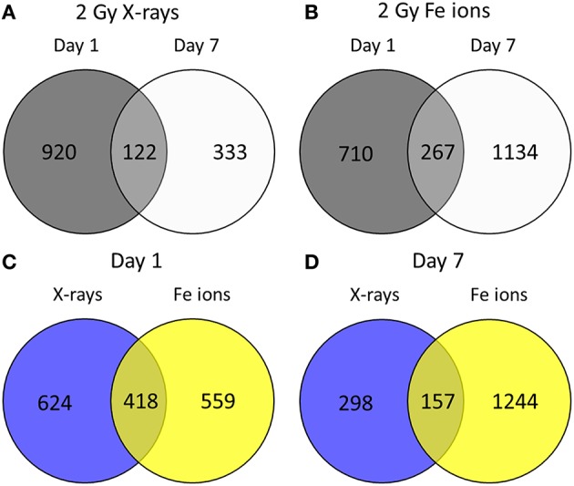
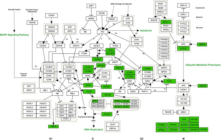
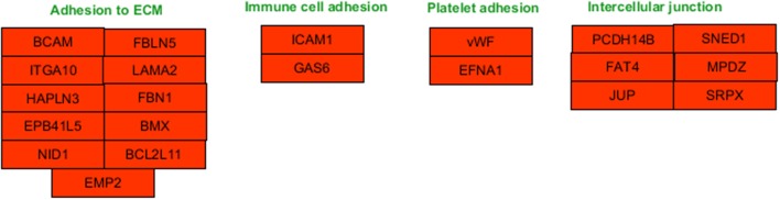
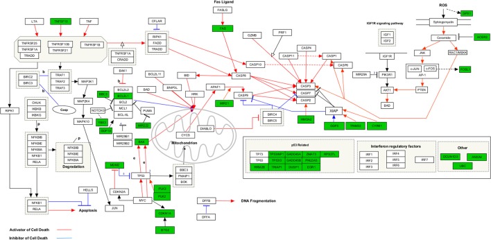
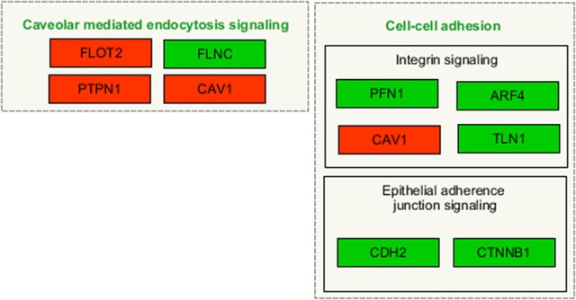
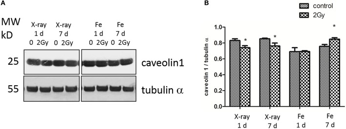
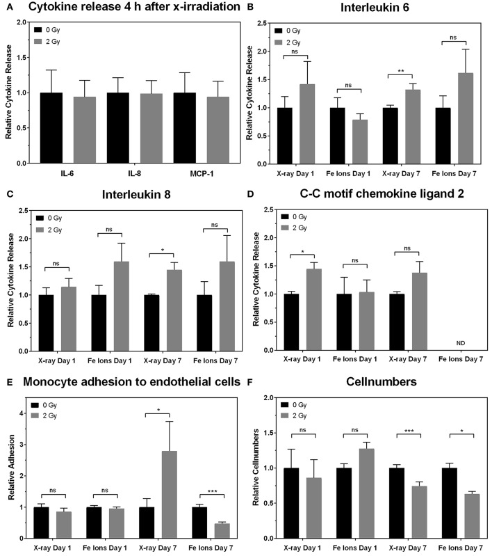
Background and Purpose: Radiotherapy is an essential tool for cancer treatment. In order to spare normal tissues and to reduce the risk of normal tissue complications, particle therapy is a method of choice. Although a large part of healthy tissues can be spared due to improved depth dose characteristics, little is known about the biological and molecular mechanisms altered after particle irradiation in healthy tissues. Elucidation of these effects is also required in the context of long term space flights, as particle radiation is the main contributor to the radiation effects observed in space. Endothelial cells (EC), forming the inner layer of all vascular structures, are especially sensitive to irradiation and, if damaged, contribute to radiation-induced cardiovascular disease. Materials and Methods: Transcriptomics, proteomics and cytokine analyses were used to compare the response of ECs irradiated or not with a single 2 Gy dose of X-rays or Fe ions measured one and 7 days post-irradiation. To support the observed inflammatory effects, monocyte adhesion on ECs was also assessed. Results: Experimental data indicate time- and radiation quality-dependent changes of the EC response to irradiation. The irradiation impact was more pronounced and longer lasting for Fe ions than for X-rays. Both radiation qualities decreased the expression of genes involved in cell-cell adhesion and enhanced the expression of proteins involved in caveolar mediated endocytosis signaling. Endothelial inflammation and adhesiveness were increased with X-rays, but decreased after Fe ion exposure. Conclusions: Fe ions induce pro-atherosclerotic processes in ECs that are different in nature and kinetics than those induced by X-rays, highlighting radiation quality-dependent differences which can be linked to the induction and progression of cardiovascular diseases (CVD). Our findings give a better understanding of the underlying processes triggered by particle irradiation in ECs, a crucial aspect for the development of protective measures for cancer patients undergoing particle therapy and for astronauts in space.

Keywords: Fe ions; X-rays; cardiovascular disease; endothelial cells; irradiation; linear energy transfer; radiotherapy.


Figures

Figure 1
Figure 1
Fe ion irradiation induces a more pronounced and persistent transcriptional response in ECs in comparison to X-ray irradiation. Venn diagrams indicate the number of differentially expressed genes after exposure to a single 2.0 Gy dose of X-rays (A) or Fe ions (B). The gene expression changes after low and high LET irradiation are compared on day 1 (C) and on day 7 (D). Changes are compared gene expression in sham irradiated cells, as described in section Materials and Methods (N = 3, n = 12).
Figure 2
Figure 2
Upregulated genes involved in cell cycle regulated processes at 7 days after a single dose of 2 Gy X-rays. Arborescence diagram indicates cell cycle pathway with the identified upregulated genes indicated in green. Pathway diagram was adapted from Wikipathways and modified with Pathvisio. Changes are shown compared to gene expression in sham irradiated cells (N = 3, n = 12).
Figure 3
Figure 3
Downregulated genes involved in cell-cell adhesion processes at 1 day after a single dose of 2 Gy X-rays. Arborescence diagram indicates cell-cell adhesion pathway with the identified downregulated genes indicated in red. Pathway diagram was adapted from Wikipathways and modified with Pathvisio. Changes are shown compared to gene expression in sham irradiated cells (N = 3, n = 12).
Figure 4
Figure 4
Upregulated genes involved in apoptosis signaling at 1 day after a single dose of 2 Gy Fe ions. Arborescence diagram indicates apoptosis pathway with the identified upregulated genes indicated in green. Pathway diagram was adapted from Wikipathways and modified with Pathvisio. Changes are shown compared to gene expression in sham irradiated cells (N = 3, n = 12).
Figure 5
Figure 5
Irradiated ECs demonstrate a larger number of differentially expressed proteins after Fe ion exposure in comparison to X-ray exposure. Venn diagrams indicate the numbers of differentially expressed proteins after exposure to a single 2.0 Gy dose of X-rays (A) or Fe ions (B). The protein expression changes after low and high LET irradiation are compared on day 1 (C) and on day 7 (D). Changes are shown compared to protein expression in sham irradiated cells, as described in section Materials and Methods (N = 3, n = 12).
Figure 6
Figure 6
Irradiated ECs differentially expressed proteins after X-ray exposure involved in caveolar-mediated endocytosis and cell-cell adhesion. Arborescence diagram indicates caveolar-mediated endocytosis signaling and cell-cell adhesion pathways with the identified upregulated genes indicated in green and downregulated genes in red. Pathway diagram was adapted from Wikipathways and modified with Pathvisio. Changes are shown compared to gene expression in sham irradiated cells (N = 3, n = 12).
Figure 7
Figure 7
X-ray irradiated ECs exhibit a persistent downregulation of caveolin-1, whereas Fe ion exposure induces a caveolin 1 upregulation. (A) Caveolin-1 (cav-1) and α-tubulin protein expression analyzed using western blot after cell exposure to a single 2.0 Gy dose fo X-ray or Fe ions. (B) Data represent the cav-1/α-tubulin ratio in control and irradiated samples after background correction and normalization to α-tubulin expression. Data show means ± SEM (N = 3, n = 12). *p < 0.05 using two-sided t-test (Welch-test).
Figure 8
Figure 8
X-ray irradiation causes EC inflammation and adhesiveness to monocytes, whilst Fe ion irradiation reduces cellular number and decreases EC adhesiveness to monocytes. The levels of IL-6, IL-8, and CCL2 secreted by EC on 4 h and day 1 plus day 7 (B–D) are shown after exposure to X-rays (n = 18–27) and Fe ions (n = 4–6). Data were normalized to cell numbers, supernatant volume, and control values. (E) The numbers of monocytes adhering to EC on day 7 are shown after exposure to X-rays (n = 90–225) and Fe ions (n = 30). Data was normalized to cell numbers and control values of sham-irradiated ECs. (F) EC numbers on day 7 using either X-rays (n = 18–27) or Fe ions (n = 4–6) are shown. (A–F). Data show mean ± SEM. ns, not significant, * p < 0.05, ** p < 0.005, *** p < 0.001 using two-sided t-test (Welch-test).

Similar articles

Cited by

References

    1. Aird W. C. (2007a). Phenotypic heterogeneity of the endothelium: I. structure, function, and mechanisms. Circ. Res. 100, 158–173. 10.1161/01.RES.0000255691.76142.4a - DOI - PubMed
    1. Aird W. C. (2007b). Phenotypic heterogeneity of the endothelium: II. representative vascular beds. Circ. Res. 100, 174–190. 10.1161/01.RES.0000255690.03436.ae - DOI - PubMed
    1. Akamatsu H., Karasawa K., Omatsu T., Isobe Y., Ogata R., Koba Y. (2014). First experience of carbon-ion radiotherapy for early breast cancer. Jpn. J. Radiol. 32, 288–295. 10.1007/s11604-014-0300-6 - DOI - PubMed
    1. Aleman B. M., Moser E. C., Nuver J., Suter T. M., Maraldo M. V., Specht L., et al. . (2014). Cardiovascular disease after cancer therapy. EJC Suppl. 12, 18–28. 10.1016/j.ejcsup.2014.03.002 - DOI - PMC - PubMed
    1. Asaithamby A., Chen D. J. (2011). Mechanism of cluster DNA damage repair in response to high-atomic number and energy particles radiation. Mutat. Res. 711, 87–99. 10.1016/j.mrfmmm.2010.11.002 - DOI - PMC - PubMed

LinkOut - more resources