Review
. 2017 Sep 25:8:499.
doi: 10.3389/fneur.2017.00499. eCollection 2017.

Electroencephalography in the Diagnosis of Genetic Generalized Epilepsy Syndromes

Affiliations
Review

Electroencephalography in the Diagnosis of Genetic Generalized Epilepsy Syndromes

Udaya Seneviratne et al. Front Neurol. .

Abstract

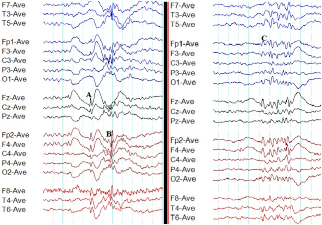
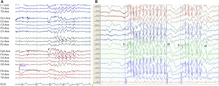
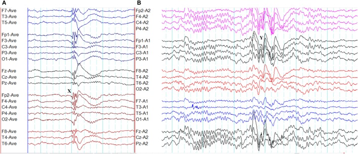
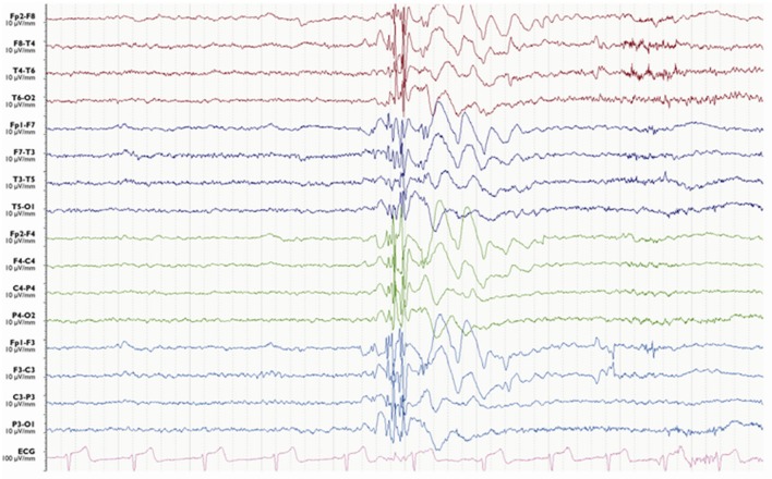
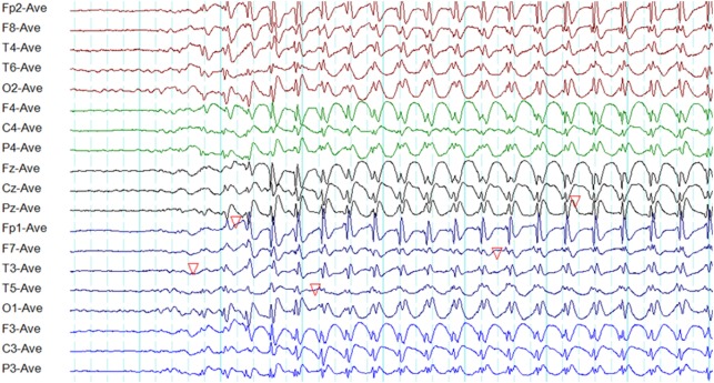
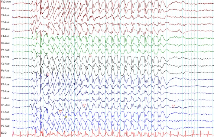
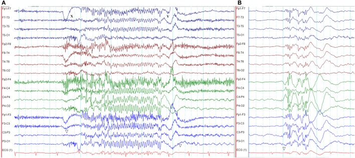
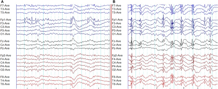
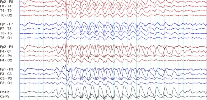
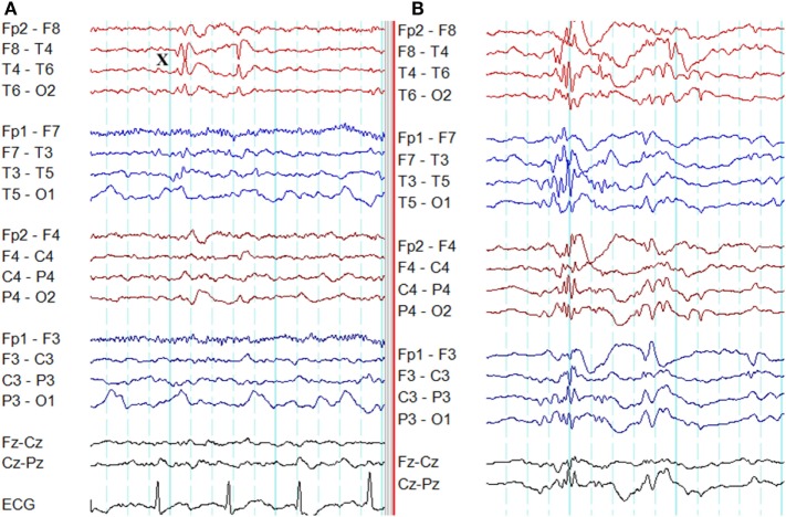
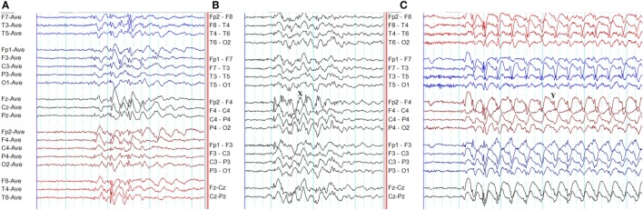
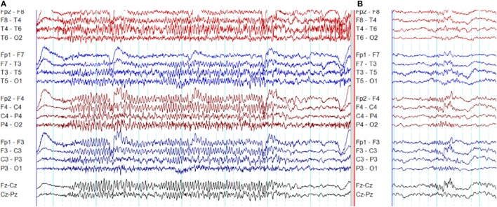
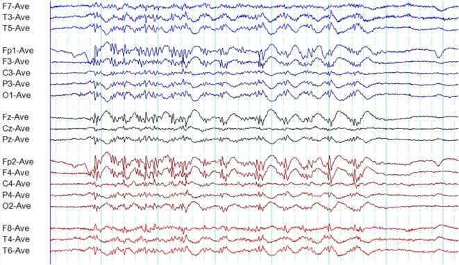
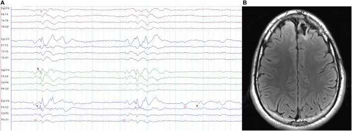
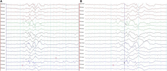
Genetic generalized epilepsy (GGE) consists of several syndromes diagnosed and classified on the basis of clinical features and electroencephalographic (EEG) abnormalities. The main EEG feature of GGE is bilateral, synchronous, symmetric, and generalized spike-wave complex. Other classic EEG abnormalities are polyspikes, epileptiform K-complexes and sleep spindles, polyspike-wave discharges, occipital intermittent rhythmic delta activity, eye-closure sensitivity, fixation-off sensitivity, and photoparoxysmal response. However, admixed with typical changes, atypical epileptiform discharges are also commonly seen in GGE. There are circadian variations of generalized epileptiform discharges. Sleep, sleep deprivation, hyperventilation, intermittent photic stimulation, eye closure, and fixation-off are often used as activation techniques to increase the diagnostic yield of EEG recordings. Reflex seizure-related EEG abnormalities can be elicited by the use of triggers such as cognitive tasks and pattern stimulation during the EEG recording in selected patients. Distinct electrographic abnormalities to help classification can be identified among different electroclinical syndromes.

Keywords: absence seizure; circadian; myoclonic seizure; photoparoxysmal response; polyspike; sleep; spike-wave; tonic-clonic seizure.


Figures

Figure 1
Figure 1
Typical interictal epileptiform discharges in genetic generalized epilepsy. Note bilateral, symmetrical, and synchronous spike-wave discharges (A), polyspike-wave discharges (B), and polyspikes (C).
Figure 2
Figure 2
Eye-closure sensitivity and fixation-off sensitivity (FOS) in genetic generalized epilepsy. (A) Generalized spike-wave and polyspike-wave discharges appear after eye closure (C) and fades away after one second indicating eye-closure sensitivity. (B) Generalized epileptiform discharges appear with eye closure (C), continues as long as eyes are closed, and disappears on eye opening (O) indicating FOS.
Figure 3
Figure 3
Epileptiform K-complexes and sleep spindles in genetic generalized epilepsy. (A) Polyspikes overlap with a K-complex at X. (B) A burst of generalized spike-wave discharges (Y) in the midst of a sleep spindle.
Figure 4
Figure 4
Electroencephalography of a myoclonic seizure. A burst of generalized polyspike-wave activity is followed a few slow waves.
Figure 5
Figure 5
Electroencephalography of a typical absence seizure. Note the paroxysm of generalized, symmetrical, synchronous, and regular 3-Hz spike-wave discharges of frontocentral maxima.
Figure 6
Figure 6
Electroencephalography (EEG) of a myoclonic absence seizure. Note the paroxysm of spike-wave discharges is similar to a typical absence seizure as illustrated in Figure 4 (time-base of this EEG = 20 s/page).
Figure 7
Figure 7
Electroencephalography of an absence seizure with eyelid myoclonia in Jeavons syndrome. (A) The absence seizure was triggered by eye closure at X. Note the paroxysm of generalized, fast polyspike activity (XY). This seizure was semiologically characterized by eyelid myoclonus, hyperextension of the neck, and unresponsiveness. (B) Interictal generalized polyspike-wave discharges during sleep recorded from the same patient.
Figure 8
Figure 8
Atypical epileptiform discharges: amplitude asymmetry. (A) Note asymmetric epileptiform discharges with higher amplitude in the left frontal region. Synchronous epileptiform discharges of low amplitude are evident on the right on careful inspection. (B) More symmetric generalized epileptiform discharges recorded from the same patient.
Figure 9
Figure 9
Atypical epileptiform discharges: focal onset and offset of paroxysms. A generalized spike-wave paroxysm in juvenile absence epilepsy. Note the focal onset and offset in the left frontal region.
Figure 10
Figure 10
Atypical epileptiform discharges: focal discharges. (A) Note focal discharges at right temporal region (X). (B) Generalized epileptiform discharges recorded from the same patient.
Figure 11
Figure 11
Atypical epileptiform discharges: abnormal morphology. (A) Waves without spikes. Note at the end of spike-wave paroxysms there are waves without preceding spikes. (B) Spikes overriding the waves. Note spikes on top of the wave at X. (C) Spikes overriding the waves. Note spikes on the descending limb of the preceding wave (Y).
Figure 12
Figure 12
Atypical epileptiform discharges: generalized paroxysmal fast rhythm. (A) A run of generalized fast activity in wakefulness. (B) Similar changes during sleep.
Figure 13
Figure 13
A disorganized (irregular) paroxysm of generalized epileptiform discharges in juvenile myoclonic epilepsy. Note this paroxysm has a mix of polyspikes and polyspike-wave discharges with varying frequency and morphology.
Figure 14
Figure 14
Pseudo bilateral synchrony in frontal lobe epilepsy. This patient presented with seizures following the surgery for left frontal brain abscess in the past. (A) In this longitudinal bipolar montage, bifrontal polyspike-wave discharges (X, Y) appear synchronous. However, focal sharp wave discharges are evident involving F3 electrode at Z. (B) The MRI demonstrating left frontal encephalomalacia.
Figure 15
Figure 15
Pseudo bilateral synchrony in frontal lobe epilepsy. (A) This average referential montage demonstrates the same activity seen in (A) of Figure 14. The discharges appear bifrontal. Note focal discharges involving F3 and C3 (time-base of the electroencephalography = 10 s/page). (B) When the time-base is expanded to 5 s/page, it becomes clear that the epileptiform discharge emerges first on the left at Fp1 and F3 (X), followed by activity on the right (Y) confirming pseudo bilateral synchrony.

Similar articles

Cited by

References

    1. Berg AT, Berkovic SF, Brodie MJ, Buchhalter J, Cross JH, van Emde Boas W, et al. Revised terminology and concepts for organization of seizures and epilepsies: report of the ILAE Commission on Classification and Terminology, 2005–2009. Epilepsia (2010) 51(4):676–85.10.1111/j.1528-1167.2010.02522.x - DOI - PubMed
    1. ILAE. Proposal for revised classification of epilepsies and epileptic syndromes. Commission on classification and terminology of the international league against epilepsy. Epilepsia (1989) 30(4):389–99.10.1111/j.1528-1157.1989.tb05316.x - DOI - PubMed
    1. Scheffer IE, Berkovic S, Capovilla G, Connolly MB, French J, Guilhoto L, et al. ILAE classification of the epilepsies: position paper of the ILAE COMMISSION for Classification and Terminology. Epilepsia (2017) 58(4):512–21.10.1111/epi.13709 - DOI - PMC - PubMed
    1. Seneviratne U, Cook M, D’Souza W. The electroencephalogram of idiopathic generalized epilepsy. Epilepsia (2012) 53(2):234–48.10.1111/j.1528-1167.2011.03344.x - DOI - PubMed
    1. Chatrian GE, Bergamini L, Dondey M, Klass DW, Lennox-Buchthal M, Petersen I. A glossary of terms most commonly used by clinical electroencephalographers. Electroencephalogr Clin Neurophysiol (1974) 37:538–48.10.1016/0013-4694(74)90099-6 - DOI - PubMed

LinkOut - more resources