. 2017 Sep 29:8:514.
doi: 10.3389/fneur.2017.00514. eCollection 2017.

Trench Foot or Non-Freezing Cold Injury As a Painful Vaso-Neuropathy: Clinical and Skin Biopsy Assessments

Affiliations

Trench Foot or Non-Freezing Cold Injury As a Painful Vaso-Neuropathy: Clinical and Skin Biopsy Assessments

Praveen Anand et al. Front Neurol. .

Abstract

Background: Trench foot, or non-freezing cold injury (NFCI), results from cold exposure of sufficient severity and duration above freezing point, with consequent sensory and vascular abnormalities which may persist for years. Based on observations of Trench foot in World War II, the condition was described as a vaso-neuropathy. While some reports have documented nerve damage after extreme cold exposure, sensory nerve fibres and vasculature have not been assessed with recent techniques in NFCI.

Objective: To assess patients with chronic sensory symptoms following cold exposure, in order to diagnose any underlying small fibre neuropathy, and provide insight into mechanisms of the persistent pain and cold hypersensitivity.

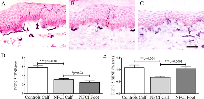
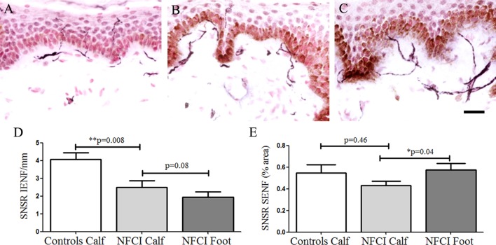
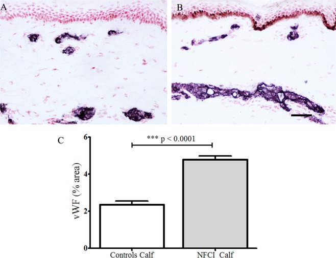
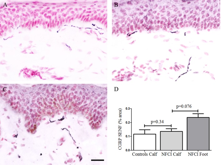
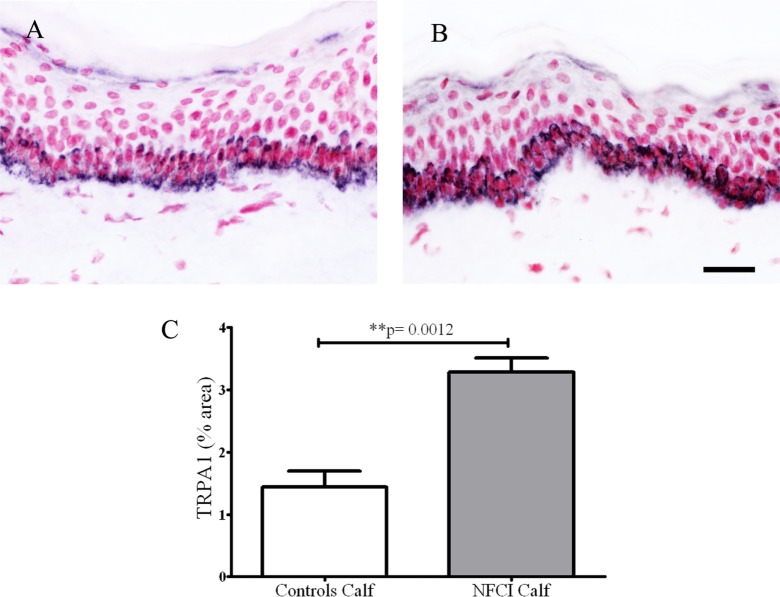
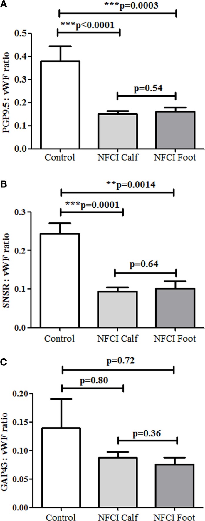
Methods: Thirty soldiers with cold exposure and persistent sensory symptoms (>4 months) were assessed with quantitative sensory testing, nerve conduction studies, and skin biopsies. Immunohistochemistry was used to assess intraepidermal (IENF) and subepidermal (SENF) nerve fibres with a range of markers, including the pan-neuronal marker protein gene product 9.5 (PGP 9.5), regenerating fibres with growth-associated protein 43 (GAP43), and nociceptor fibres with transient receptor potential cation channel subfamily V member 1 (TRPV1), sensory neuron-specific receptor (SNSR), and calcitonin gene-related peptide (CGRP). von Willebrand factor (vWF), endothelial nitric oxide synthase (eNOS), and vascular endothelial growth factor (VEGF) were used for assessing blood vessels, and transient receptor potential cation channel, subfamily A member 1 (TRPA1) and P2X purinoceptor 7 (P2X7) for keratinocytes, which regulate nociceptors via release of nerve growth factor.

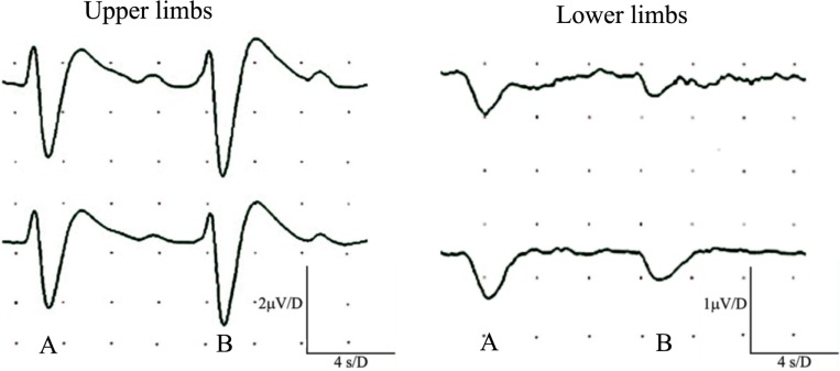
Results: Clinical examination showed pinprick sensation was abnormal in the feet of 20 patients (67%), and between 67 and 83% had abnormalities of thermal thresholds to the different modalities. 7 patients (23%) showed reduced sensory action potential amplitude of plantar nerves. 27 patients (90%) had decreased calf skin PGP 9.5 IENF (p < 0.0001), the remaining 3 patients had decreased nerve markers in subepidermis or foot skin. There were marked increases of all vascular markers (for vWF in calf skin, p < 0.0001), and increased sensory or regenerating SENF (for calf skin, GAP43, p = 0.002). TRPA1 (p = 0.0012) and P2X7 (p < 0.0001) were increased in basal keratinocytes.

Conclusion: A range of skin biopsy markers and plantar nerve conduction studies are useful objective assessments for the diagnosis of peripheral neuropathy in NFCI. Our results suggest that an increase in blood vessels following tissue ischaemia/hypoxia could be associated with disproportionate and abnormal nerve fibres (irritable nociceptors), and may lead to NFCI as a "painful vaso-neuropathy."

Keywords: nerve conduction study; neuropathy; non-freezing cold injury; pain; skin biopsy; trench foot.


Figures

Figure 1
Figure 1
Nerve conduction studies in patients with non-freezing cold injury. Sensory nerve action potential of the plantar nerves. (A) Normal, (B) reduced amplitude, and (C) absent.
Figure 2
Figure 2
Simultaneous recordings of normal sympathetic skin responses (SSR) from both hands and feet. SSR evoked by light, sudden unexpected tactile stimulus (A), and a sudden inspiratory gasp (B).
Figure 3
Figure 3
Protein gene product 9.5 (PGP 9.5) immunoreactivity in skin. PGP 9.5 staining in control calf skin (A), in non-freezing cold injury (NFCI) calf skin (B), and in NFCI foot skin (C); scale bar = 50 µm; bar charts of the PGP 9.5 intraepidermal nerve fibre (IENF)/mm (D); bar charts of PGP 9.5 subepidermal nerve fibre (SENF) (% area) (E).
Figure 4
Figure 4
Transient receptor potential cation channel subfamily V member 1 (TRPV1) immunoreactivity in skin. TRPV1 staining in control calf skin (A), in non-freezing cold injury (NFCI) calf skin (B); scale bar = 50 µm; bar charts of the TRPV1 intraepidermal nerve fibre (IENF) fibres/mm (C); bar charts of the image analysis of TRPV1 subepidermal nerve fibre (SENF) (% area) (D).
Figure 5
Figure 5
Sensory neuron-specific receptor (SNSR) immunoreactivity in skin. SNSR staining in control calf skin (A), in non-freezing cold injury (NFCI) calf skin (B), and in NFCI foot skin (C); scale bar = 50 µm; bar charts of the SNSR intraepidermal nerve fibre (IENF) fibres/mm (D); bar charts of the image analysis of the SNSR subepidermal nerve fibre (SENF) (% area) (E).
Figure 6
Figure 6
Growth-associated protein 43 (GAP43) immunoreactivity in skin. GAP43 staining in control calf skin (A), in non-freezing cold injury (NFCI) calf skin (B), and in NFCI foot skin (C); scale bar = 50 µm; bar charts of the GAP43 intraepidermal nerve fibre (IENF) fibres/mm (D); bar charts of GAP43 subepidermal nerve fibre (SENF) (% area) (E).
Figure 7
Figure 7
von Willebrand Factor (vWF) immunoreactivity in skin. vWF staining in control calf skin (A) and in non-freezing cold injury (NFCI) calf skin (B); scale bar = 100 µm; bar chart of the image analysis of the vWF immunoreactivity (C).
Figure 8
Figure 8
Vascular endothelial growth factor (VEGF) immunoreactivity in skin. VEGF staining in control calf skin (A) and in non-freezing cold injury (NFCI) calf skin (B); scale bar = 100 µm; bar chart of the image analysis of the VEGF immunoreactivity (C).
Figure 9
Figure 9
Endothelial nitric oxide synthase (eNOS) immunoreactivity in skin. eNOS staining in control calf skin (A) and in non-freezing cold injury (NFCI) calf skin (B); scale bar = 100 µm; bar chart of the image analysis of the eNOS immunoreactivity (C).
Figure 10
Figure 10
Calcitonin gene-related peptide (CGRP) immunoreactivity in skin. CGRP staining in control calf skin (A), in non-freezing cold injury (NFCI) calf skin (B), and in NFCI foot skin (C); scale bar = 50 µm. Bar chart of the image analysis of the CGRP subepidermal nerve fibre (SENF) (% area) (D).
Figure 11
Figure 11
Transient receptor potential cation channel, subfamily A member 1 (TRPA1) immunoreactivity in skin. Basal keratinocyte cell staining with TRPA1 in control calf skin (A) or non-freezing cold injury (NFCI) calf skin (B); scale bar = 50 µm. Bar chart of the image analysis of the TRPA1 immunoreactivity (C).
Figure 12
Figure 12
P2X purinoceptor 7 (P2X7) receptor immunoreactivity in skin. Keratinocyte and nerve fibre (arrowed) staining in control calf skin (A) and non-freezing cold injury (NFCI) calf skin (B); bar chart of the image analysis of P2X7 receptor epidermal immunoreactivity in calf skin (C); P2X7 receptor immunoreactive nerve fibres (arrowed) in the subepidermal region (D), and suprabasal keratinocyte staining (arrowheads) to the top of the epidermis, in NFCI foot skin (E); P2X7 receptor nerve fibre bundle in deep dermis of foot skin (F); scale bar = 50 µm.
Figure 13
Figure 13
Neuronal and vascular colocalisation staining in skin. Association of protein gene product 9.5 (A,B) or sensory neuron-specific receptor (C,D) neuronal staining (black), with the blood vessel endothelial cell marker cluster of differentiation 31 (red), in the subepidermis of non-freezing cold injury foot skin; scale bar = 50 µm.
Figure 14
Figure 14
Ratios of subepidermal nerve fibres (SENFs) to von Willebrand factor (vWF) in non-freezing cold injury (NFCI) skin. Ratios of nerve markers to vWF, (A) protein gene product 9.5 (PGP 9.5) (PGP 9.5:vWF) in control and NFCI calf skin, (B) sensory neuron-specific receptor (SNSR) (SNSR:vWF), and (C) growth-associated protein 43 (GAP43) (GAP43:vWF).

Similar articles

Cited by

References

    1. Lorrain Smith J, Ritchie J, Dawson J. Clinical and experimental observations on the pathology of trench frostbite. J Pathol Bacteriol (1915) 20(1):159–90.10.1002/path.1700200113 - DOI
    1. Atenstaedt RL. Trench foot: the medical response in the first World War 1914–18. Wilderness Environ Med (2006) 17(4):282–9.10.1580/06-WEME-LH-027R.1 - DOI - PubMed
    1. Hughes B. The causes and prevention of trench foot. Br Med J (1916) 1(2890):712–4.10.1136/bmj.1.2890.712 - DOI - PMC - PubMed
    1. Ungley CC, Blackwood W. Peripheral vasoneuropathy after chilling. “Immersion foot and immersion hand”. Lancet (1942) 240(6216):447–51.10.1016/S0140-6736(00)58135-5 - DOI
    1. Ungley CC, Channell GD, Richards RL. The immersion foot syndrome. Br J Surg (1945) 33(129):17–31.10.1002/bjs.18003312903 - DOI - PubMed