Review
. 2018:247:87-114.
doi: 10.1007/164_2017_58.

Delta Opioid Receptor Expression and Function in Primary Afferent Somatosensory Neurons

Affiliations
Review

Delta Opioid Receptor Expression and Function in Primary Afferent Somatosensory Neurons

Amaury François et al. Handb Exp Pharmacol. 2018.

Abstract

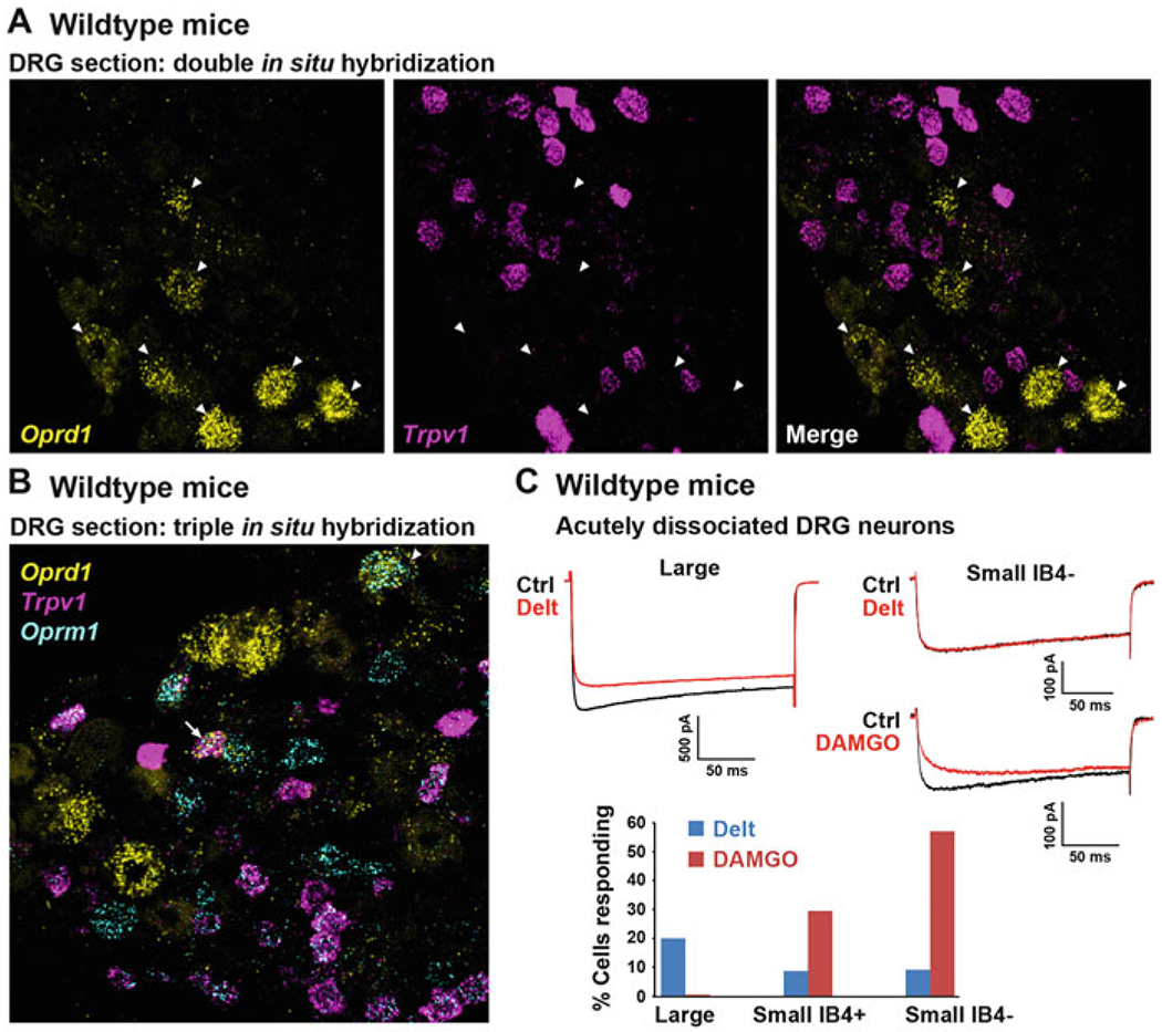
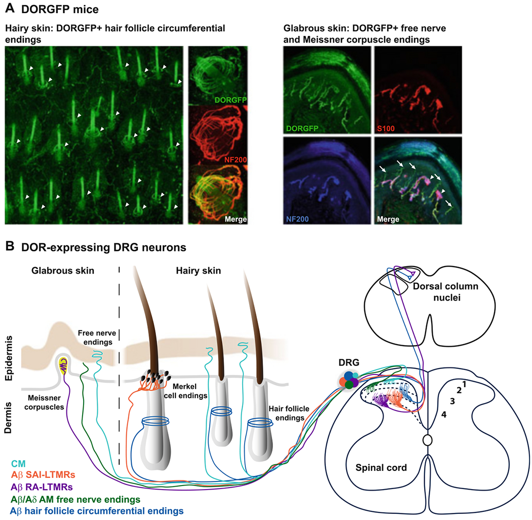
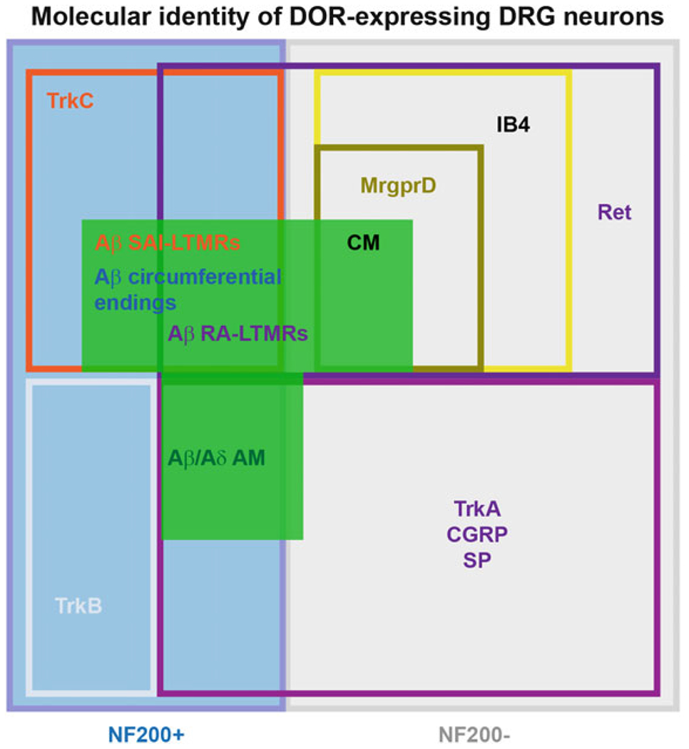
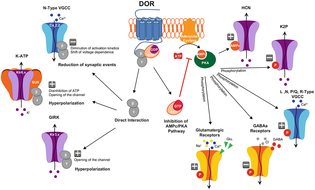
The functional diversity of primary afferent neurons of the dorsal root ganglia (DRG) generates a variety of qualitatively and quantitatively distinct somatosensory experiences, from shooting pain to pleasant touch. In recent years, the identification of dozens of genetic markers specifically expressed by subpopulations of DRG neurons has dramatically improved our understanding of this diversity and provided the tools to manipulate their activity and uncover their molecular identity and function. Opioid receptors have long been known to be expressed by discrete populations of DRG neurons, in which they regulate cell excitability and neurotransmitter release. We review recent insights into the identity of the DRG neurons that express the delta opioid receptor (DOR) and the ion channel mechanisms that DOR engages in these cells to regulate sensory input. We highlight recent findings derived from DORGFP reporter mice and from in situ hybridization and RNA sequencing studies in wild-type mice that revealed DOR presence in cutaneous mechanosensory afferents eliciting touch and implicated in tactile allodynia. Mechanistically, we describe how DOR modulates opening of voltage-gated calcium channels (VGCCs) to control glutamatergic neurotransmission between somatosensory neurons and postsynaptic neurons in the spinal cord dorsal horn. We additionally discuss other potential signaling mechanisms, including those involving potassium channels, which DOR may engage to fine tune somatosensation. We conclude by discussing how this knowledge may explain the analgesic properties of DOR agonists against mechanical pain and uncovers an unanticipated specialized function for DOR in cutaneous mechanosensation.

Keywords: Delta opioid receptor; Excitability; Ion channels; Mechanosensation; Neuroanatomy; Neurotransmitter release; Pain; Primary afferent dorsal root ganglion neurons; Touch.


Figures

Fig. 1
Fig. 1
DOR is predominantly expressed by large-diameter DRG neurons, not by TRPV1+ and MOR+ small-diameter C nociceptors. (a) Single mRNA molecule labeling in DRG sections from wild-type mice reveals that DOR (Oprd1 gene) and TRPV1 are expressed by different populations of DRG neurons. Arrowheads indicate DOR+ neurons. (b) The great majority of TRPV1+ peptidergic C nociceptors co-express MOR (Oprm1 gene). MOR is also found in other DRG neuron types, including large-diameter neurons in which it occasionally co-occurs with DOR (arrowhead). The arrow shows a rare example of a C nociceptor co-expressing MOR, TRPV1, and DOR. (c) Consistent with histological data, electrophysiological recordings show that the DOR agonist deltorphin II (Delt), in contrast to the MOR agonist DAMGO, predominantly inhibits calcium currents in large-diameter DRG neurons from wild-type mice (modified from Bardoni et al. 2014). Images in a and b were provided by Dong Wang, Scherrer laboratory
Fig. 2
Fig. 2
DOR is expressed by several classes of cutaneous mechanosensitive neurons. (a) DORGFP mice reveal DOR expression at the peripheral terminals of A low-threshold mechanoreceptors (LTMRs) forming circumferential endings around hair follicles (arrowheads in left panel). DOR is also expressed by DRG neurons innervating the glabrous skin, particularly by C nonpeptidergic nociceptors that co-express MrgprD and end as free nerve endings in the stratum granulosum (arrows in right panel) and by A LTMRs forming Meissner corpuscles (arrowheads in right panel). Modified from Bardoni et al. (2014). (b) Schematic summarizing the peripheral and central projection patterns of DOR+ cutaneous DRG neurons. CM C mechanonociceptors, SA slowly adapting, RA rapidly adapting
Fig. 3
Fig. 3
Functional and genetic diversity of A LTMRs and nociceptors expressing DOR. Multiple classes of molecularly heterogeneous DRG neurons express DOR. These different populations include myelinated nociceptors that express TrkA, as well as A LTMRs expressing other neurotrophin receptors such as Ret and TrkC. Ret is also expressed by the C nociceptors that express DOR, which correspond to a subpopulation of IB4-binding MrgprD+ mechanosensitive afferents
Fig. 4
Fig. 4
Putative ion channel mechanisms engaged by DOR in DRG neurons. Binding of endogenous or exogenous DOR agonists activates G proteins. Subsequent separation of the G protein subunits triggers distinct signaling cascades: on the left, direct interaction of Gβγ with N-Type VGCCs leads to reduced calcium current amplitude and decreased neurotransmitter release, while direct interaction with GIRK and/or KATP channels increases potassium conductance, resulting in membrane hyperpolarization. On the right, inhibition of adenylate cyclase by Gαi/o reduces cAMP production and PKA activation, which alters the phosphorylation states of different receptors and ion channels and leads to decreased excitability

Similar articles

Cited by

References

    1. Abraira VE, Ginty DD (2013) The sensory neurons of touch. Neuron 79:618–639 - PMC - PubMed
    1. Acosta CG, Lopez HS (1999) Delta opioid receptor modulation of several voltage-dependent Ca(2+) currents in rat sensory neurons. J Neurosci 19:8337–8348 - PMC - PubMed
    1. Afify EA, Khedr MM, Omar AG, Nasser SA (2013) The involvement of K(ATP) channels in morphine-induced antinociception and hepatic oxidative stress in acute and inflammatory pain in rats. Fundam Clin Pharmacol 27:623–631 - PubMed
    1. Aguilar-Bryan L et al. (1998) Toward understanding the assembly and structure of KATP channels. Physiol Rev 78:227–245 - PubMed
    1. Al-Hasani R, Bruchas MR (2011) Molecular mechanisms of opioid receptor-dependent signaling and behavior. Anesthesiology 115:1363–1381 - PMC - PubMed

Publication types

Substances

LinkOut - more resources