. 2018 Feb;33(2):433-447.
doi: 10.1007/s12640-017-9812-z. Epub 2017 Oct 9.

Methamphetamine Augments Concurrent Astrocyte Mitochondrial Stress, Oxidative Burden, and Antioxidant Capacity: Tipping the Balance in HIV-Associated Neurodegeneration

Affiliations

Methamphetamine Augments Concurrent Astrocyte Mitochondrial Stress, Oxidative Burden, and Antioxidant Capacity: Tipping the Balance in HIV-Associated Neurodegeneration

Kathleen Borgmann et al. Neurotox Res. 2018 Feb.

Abstract

Methamphetamine (METH) use, with and without human immunodeficiency virus (HIV)-1 comorbidity, exacerbates neurocognitive decline. Oxidative stress is a probable neurotoxic mechanism during HIV-1 central nervous system infection and METH abuse, as viral proteins, antiretroviral therapy and METH have each been shown to induce mitochondrial dysfunction. However, the mechanisms regulating mitochondrial homeostasis and overall oxidative burden in astrocytes are not well understood in the context of HIV-1 infection and METH abuse. Here, we report METH-mediated dysregulation of astrocyte mitochondrial morphology and function during prolonged exposure to low levels of METH. Mitochondria became larger and more rod shaped with METH when assessed by machine learning, segmentation analyses. These changes may be mediated by elevated mitofusin expression coupled with inhibitory phosphorylation of dynamin-related protein-1, which regulate mitochondrial fusion and fission, respectively. While METH decreased oxygen consumption and ATP levels during acute exposure, chronic treatment of 1 to 2 weeks significantly enhanced both when tested in the absence of METH. Together, these changes significantly increased not only expression of antioxidant proteins, augmenting the astrocyte's oxidative capacity, but also oxidative damage. We propose that targeting astrocytes to reduce their overall oxidative burden and expand their antioxidant capacity could ultimately tip the balance from neurotoxicity towards neuroprotection.

Keywords: Astroglia; Dynamin-related protein; Extracellular flux; Machine learning; Mitochondria; Mitofusin; Neurotoxicity; Oxidative stress.


Figures

Fig. 1
Fig. 1. Astrocyte mitochondria become enlarged with prolonged exposure to METH
Human astrocytes were exposed to 0 nM (a, Control), 50 nM (b), 5 μM (c), or 500 μM (d) METH for one week. Mitochondria were then fluorescently labeled with Mitotracker red (MTR, red) and nuclei with Hoechst (blue). Live cultures were imaged at an original magnification of 400X. Total mitochondria were segmented into morphological types including punctate (e, i), rod (f, j) large spots (g, k) and networks (h, l) with Weka machine learning in Fiji, ImageJ (see supplementary Fig. 1 for example layers). The area occupied by mitochondria of each type (e, f, g, h) and fold change in average size (i, j, k, l) were calculated based on the analysis of segmented layers in each of 10–15 micrographs per condition. Representative images of each mitochondrial morphology are shown, below representative images from each treatment. Statistical significance was determined by one-way ANOVA followed by Dunnett’s multiple comparisons test with GraphPad Prism (V7) and error bars are standard error of the mean (SEM). *P<0.05, **P<0.01, ***P<0.001
Fig. 2
Fig. 2. METH increases mitochondrial regulatory protein levels and dysregulates their activity
Primary human astrocyte cultures were treated with METH (50 nM and 5 μM) for one week. Levels of cytochrome c oxidase (COX) IV (a, e), mitofusin (MFN) (b, f), dynamin-related protein 1 (Drp-1) (c, g) and microtubule associated protein light chain 3 (LC3) (d, h) were evaluated by real-time PCR and immunoblotting or WES, respectively. Graphs are cumulative average expression level in 3 to 6 separate astrocyte donors and representative blots are shown. Statistical significance was determined by one-way ANOVA followed by Dunnett’s multiple comparisons test (a–d) or student’s test (e–g) with GraphPad Prism (V7). Error bars are SEM. Hatched lines indicate blot cropping of non-adjacent bands. *P<0.05, **P<0.01
Fig. 3
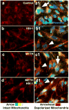
Fig. 3. Mitochondrial permeability transition pores (mPTP) open during HIV-1 exposure
Astrocyte cultures were treated with HIV-1JR-FL (10 ng/ml HIV-1 p24, b/b1), METH (500 μM, c/c1), or both (d/d1) for 24 hours. Control (a/a1) and treated astrocytes were then fluorescently labeled with Mitotracker Red® (MTR, red), calcein-AM (green, when sequestered in mitochondria) and the nuclear stain Hoechst (blue). Examples of astrocytes with closed mPTP are indicated by red/green (yellow) mitochondrial fluorescence (arrows). Open mPTP are visualized by decreased mitochondrial green fluorescence (arrowheads), as calcein is quenched when released into the cytoplasm by cobalt chloride, suggesting mitochondrial dysfunction. Original magnification 200X
Fig. 4
Fig. 4. Prolonged METH increases oxygen consumption and ATP levels in primary human astrocytes
Since TAAR1 is a known astrocyte receptor for METH, astrocytes were pretreated with the selective TAAR1 antagonist EPPTB (20 μM) for one hour (a). Oxygen consumption was measured by extracellular flux assay (Seahorse) with the sequential injection of 100 μM and 500 μM METH over the course of three hours. Oxygen consumption rates (OCR) were normalized to control (0 μM) and EPPTB pretreated rates, respectively on a per well basis. Oligomycin, an ATP synthase inhibitor, was used as a positive control for decreased OCR. Bars represent the average OCR in seven separate astrocyte cultures +/− SEM. In a separate experiment ATP levels were measured three hours post-METH treatment (50 nM-500 μM) by luciferase assay and compared to control (0 nM) levels (b). Bars represent the average fold change in relative light units (RLU) in three separate astrocyte cultures +/− SEM. In another paradigm, astrocytes were treated with 50 nM METH for one week and OCR (c) and ATP levels (d) were assayed following METH withdrawl. Bars represent the average OCR or fold change in ATP in two independent astrocyte cultures +/− SEM. Statistical significance was determined by one-way ANOVA followed by Dunnett’s multiple comparisons test (a–b) or student’s test (c–d) with GraphPad Prism (V7). **P<0.01, ***P<0.001
Fig. 5
Fig. 5. Chronic METH exposure increases the antioxidant capacity and oxidative burden of human astrocytes
To examine the effects of prolonged METH exposure, astrocytes were treated with METH between 50 nM and 500 μM for two weeks. Untreated control (0 nM) astrocytes were maintained in parallel. Oxygen consumption rate (OCR) was measured in the absence of METH by extracellular flux Seahorse assay (a). Antioxidant protein expression was measured by real-time PCR. The average fold change in glutathione reductase (b), superoxide dismutase (SOD) 1(c), SOD2 (d) and catalase (e) expression levels as compared to control (0 nM) are shown. As measure of oxidative balance, levels of malondialdehyde (MDA)-modified proteins and SOD1 were evaluated in total cell lysates by immunoblotting in parallel. GAPDH levels were evaluated as housekeeping and loading controls, respectively. Panels are representative experiments repeated in two independent astrocyte donors. Significance was determined with one-way ANOVA followed by Dunnett’s multiple comparisons test with GraphPad Prism (V7). **P<0.01, ***P<0.001
Fig. 6
Fig. 6. METH disturbs the delicate Yin & Yang of oxidative burden and antioxidant capacity in human astrocytes
Prolonged METH exposure induces changes in mitochondrial morphology and size in astrocytes (Fig. 1). Mitochondria become larger and rod shaped. Direct and indirect effects of METH on intracellular signaling and the electron transport chain likely contribute to this phenomenon. METH increases astrocyte mitofusin (MFN) levels, which may promote mitochondrial fusion and networking. In parallel, METH induces inhibitory phosphorylation of dynamin-related protein-1 (Drp-1) at serine 637, which may decrease mitochondrial fission (Fig. 2). Together these changes in mitochondrial regulatory proteins may disturb mitochondrial homeostasis, morphology and oxidative stress. HIV-1 induces mitochondrial permeability transition pore (mPTP) opening (Fig. 3), a measure of mitochondrial stress and dysfunction, while acute METH exposure impairs mitochondrial respiration. Prolonged METH exposure augments oxygen consumption (O2) and ATP availability (Fig. 4), suggesting an overall increase in astrocyte mitochondria. METH-mediated oxidative stress promotes the expression of antioxidant response element (ARE) regulated genes increasing antioxidant levels in astrocytes (Fig. 5). Since neurons are highly dependent on astrocytes for antioxidant support, neurotoxicity successfully prevented if the antioxidant capacity of METH-exposed astrocytes is greater than the oxidative burden of astrocyte and adjacent neurons combined. Astrocyte directed therapy could tip the balance towards increasing antioxidant capacity while reducing oxidative burden.

Similar articles

Cited by

References

    1. Anton F, Dittmar G, Langer T, Escobar-Henriques M. Two deubiquitylases act on mitofusin and regulate mitochondrial fusion along independent pathways. Mol Cell. 2013;49:487–498. doi: 10.1016/j.molcel.2012.12.003. - DOI - PubMed
    1. Arganda-Carreras I, Kaynig V, Rueden C, Eliceiri KW, Schindelin J, Cardona A, Seung HS. Trainable Weka Segmentation: a machine learning tool for microscopy pixel classification. Bioinformatics. 2017 doi: 10.1093/bioinformatics/btx180. - DOI - PubMed
    1. Avdoshina V, Fields JA, Castellano P, Dedoni S, Palchik G, Trejo M, Adame A, Rockenstein E, Eugenin E, Masliah E, Mocchetti I. The HIV Protein gp120 Alters Mitochondrial Dynamics in Neurons. Neurotox Res. 2016;29:583–593. doi: 10.1007/s12640-016-9608-6. - DOI - PMC - PubMed
    1. Barnham KJ, Masters CL, Bush AI. Neurodegenerative diseases and oxidative stress. Nat Rev Drug Discov. 2004;3:205–214. doi: 10.1038/nrd1330. - DOI - PubMed
    1. Baxter PS, Hardingham GE. Adaptive regulation of the brain’s antioxidant defences by neurons and astrocytes. Free Radic Biol Med. 2016;100:147–152. doi: 10.1016/j.freeradbiomed.2016.06.027. - DOI - PMC - PubMed

Publication types

MeSH terms