

Age-associated dysregulation of protein metabolism in the mammalian oocyte
- PMID: 28994181
- PMCID: PMC5676066
- DOI: 10.1111/acel.12676
Age-associated dysregulation of protein metabolism in the mammalian oocyte
Abstract
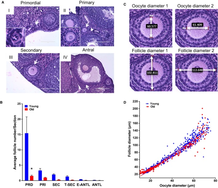
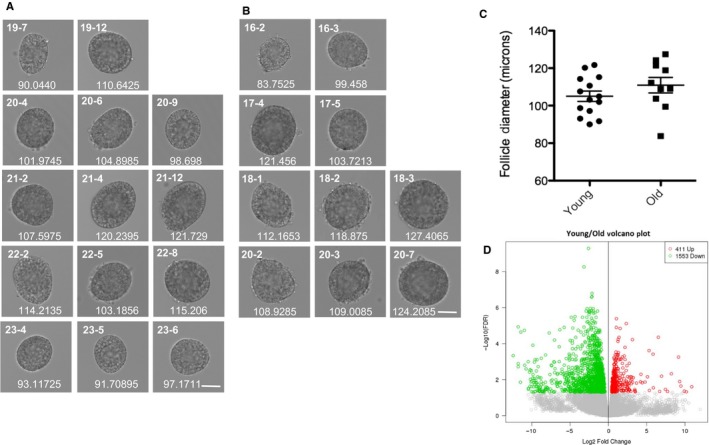
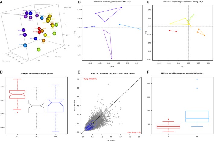
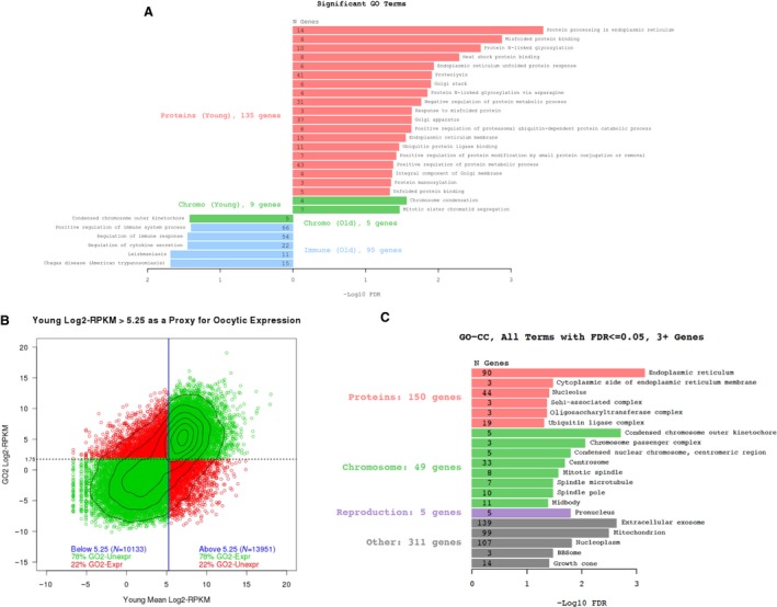
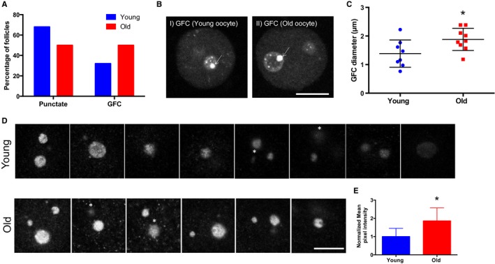
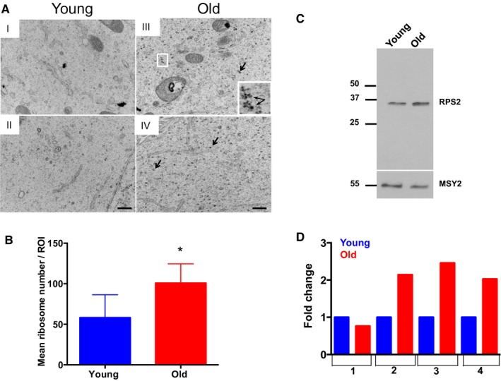
Reproductive aging is characterized by a marked decline in oocyte quality that contributes to infertility, miscarriages, and birth defects. This decline is multifactorial, and the underlying mechanisms are under active investigation. Here, we performed RNA-Seq on individual growing follicles from reproductively young and old mice to identify age-dependent functions in oocytes. This unbiased approach revealed genes involved in cellular processes known to change with age, including mitochondrial function and meiotic chromosome segregation, but also uncovered previously unappreciated categories of genes related to proteostasis and organelles required for protein metabolism. We further validated our RNA-Seq data by comparing nucleolar structure and function in oocytes from reproductively young and old mice, as this organelle is central for protein production. We examined key nucleolar markers, including upstream binding transcription factor (UBTF), an RNA polymerase I cofactor, and fibrillarin, an rRNA methyltransferase. In oocytes from mice of advanced reproductive age, UBTF was primarily expressed in giant fibrillar centers (GFCs), structures associated with high levels of rDNA transcription, and fibrillarin expression was increased ~2-fold. At the ultrastructural level, oocyte nucleoli from reproductively old mice had correspondingly more prominent fibrillar centers and dense fibrillar centers relative to young controls and more ribosomes were found in the cytoplasm. Taken together, our findings are significant because the growing oocyte is one of the most translationally active cells in the body and must accumulate high-quality maternally derived proteins to support subsequent embryo development. Thus, perturbations in protein metabolism are likely to have a profound impact on gamete health.
Keywords: folliculogenesis; nucleolus; oogenesis; proteostasis; reproductive aging; ribosome.
© 2017 The Authors. Aging Cell published by the Anatomical Society and John Wiley & Sons Ltd.
Figures
Comment in
-
Mammalian oogenesis and female reproductive aging.Aging (Albany NY). 2018 Feb 5;10(2):162-163. doi: 10.18632/aging.101381. Aging (Albany NY). 2018. PMID: 29410392 Free PMC article. No abstract available.
References
-
- Ben‐Meir A, Burstein E, Borrego‐Alvarez A, Chong J, Wong E, Yavorska T, Naranian T, Chi M, Wang Y, Bentov Y, Alexis J, Meriano J, Sung HK, Gasser DL, Moley KH, Hekimi S, Casper RF, Jurisicova A (2015) Coenzyme Q10 restores oocyte mitochondrial function and fertility during reproductive aging. Aging Cell 14, 887–895. - PMC - PubMed
-
- Bristol‐Gould SK, Kreeger PK, Selkirk CG, Kilen SM, Cook RW, Kipp JL, Shea LD, Mayo KE, Woodruff TK (2006) Postnatal regulation of germ cells by activin: the establishment of the initial follicle pool. Dev. Biol. 298, 132–148. - PubMed
Publication types
MeSH terms
Substances
Grants and funding
LinkOut - more resources
Full Text Sources
Other Literature Sources
