Classification of Lacrimal Punctal Stenosis and Its Related Histopathological Feature in Patients with Epiphora
- PMID: 28994268
- PMCID: PMC5636712
- DOI: 10.3341/kjo.2016.0129
Classification of Lacrimal Punctal Stenosis and Its Related Histopathological Feature in Patients with Epiphora
Abstract
Purpose: To evaluate the classification of punctal stenosis based on the shape of the external punctum, clinical characteristics and histopathologic features.
Methods: Patients who experienced tearing and were diagnosed with punctal stenosis were evaluated in this study. Punctal stenosis was classified according to the shape of the lower external punctum, which included membranous type, slit type, horseshoe type, and pinpoint type. Tear meniscus height, 2% fluorescein dye disappearance test and lacrimal pathway irrigation were measured or performed. For treatment, a punctal snip operation and silicone tube placement were performed, and the peripunctal histopathological findings were evaluated.
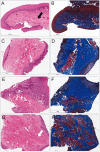
Results: Punctal stenosis was classified into four types: membranous type (17 eyes, 21.5%), slit type (11 eyes, 13.9%), horseshoe type (25 eyes, 31.6%), and pinpoint type (26 eyes, 32.9%). The tear meniscus was significantly higher, and the 2% fluorescein dye disappeared significantly more slowly in the punctal stenosis group. However, correlation of the tear meniscus height and 2% fluorescein dye disappearance test with the punctum shape was not statistically significant. A history of previous chemotherapy was significantly associated with the occurrence of punctal stenosis, especially the membranous type (p < 0.05). Histopathologic evaluation of the punctum showed differences between the punctum types. Pinpoint puncta exhibited a high density of muscle fibers, while they were faintly visible in the membranous type.
Conclusions: Acquired punctal stenosis has various shapes, and the major types of stenotic puncta exhibited unique histopathologic features. Punctal stenosis and its pathophysiology may be related to multiple factors, such as age and systemic 5-fluorouracil chemotherapy history.
Keywords: Histopathological; Lacrimal apparatus; Lacrimal apparatus diseases.
© 2017 The Korean Ophthalmological Society
Conflict of interest statement
No potential conflict of interest relevant to this article was reported.
Figures


References
-
- Kakizaki H, Takahashi Y, Iwaki M, et al. Punctal and canalicular anatomy: implications for canalicular occlusion in severe dry eye. Am J Ophthalmol. 2012;153:229–237. - PubMed
-
- Lipham WJ, Tawfik HA, Dutton JJ. A histologic analysis and three-dimensional reconstruction of the muscle of Riolan. Ophthal Plast Reconstr Surg. 2002;18:93–98. - PubMed
-
- Kashkouli MB, Beigi B, Astbury N. Acquired external punctal stenosis: surgical management and long-term follow-up. Orbit. 2005;24:73–78. - PubMed
-
- Kristan RW. Treatment of lacrimal punctal stenosis with a one-snip canaliculotomy and temporary punctal plugs. Arch Ophthalmol. 1988;106:878–879. - PubMed
MeSH terms
LinkOut - more resources
Full Text Sources
Other Literature Sources

