

Three-dimensional Imaging and Analysis of Mitochondria within Human Intraepidermal Nerve Fibers
- PMID: 28994751
- PMCID: PMC5752341
- DOI: 10.3791/53369
Three-dimensional Imaging and Analysis of Mitochondria within Human Intraepidermal Nerve Fibers
Abstract
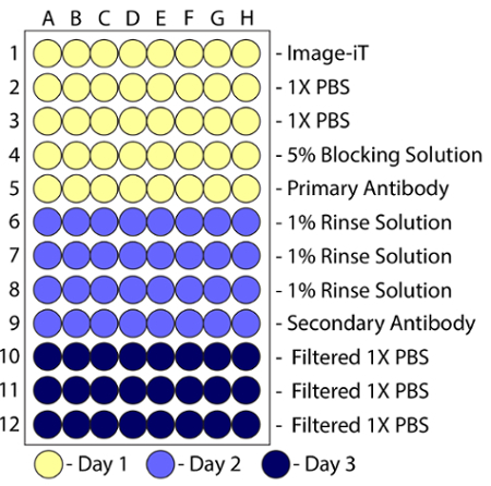
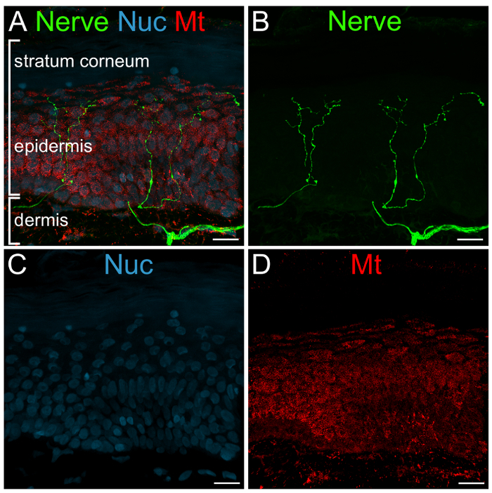
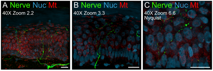
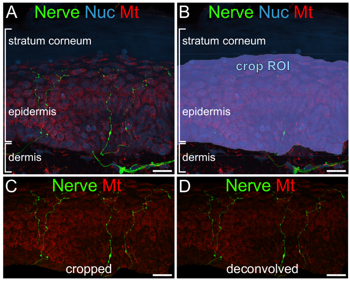
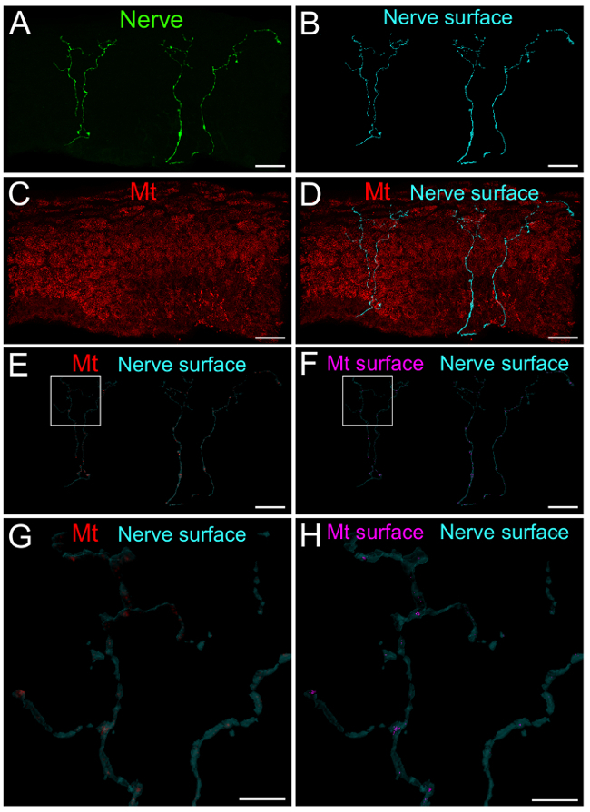
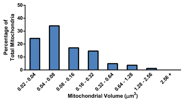
The goal of this protocol is to study mitochondria within intraepidermal nerve fibers. Therefore, 3D imaging and analysis techniques were developed to isolate nerve-specific mitochondria and evaluate disease-induced alterations of mitochondria in the distal tip of sensory nerves. The protocol combines fluorescence immunohistochemistry, confocal microscopy and 3D image analysis techniques to visualize and quantify nerve-specific mitochondria. Detailed parameters are defined throughout the procedures in order to provide a concrete example of how to use these techniques to isolate nerve-specific mitochondria. Antibodies were used to label nerve and mitochondrial signals within tissue sections of skin punch biopsies, which was followed by indirect immunofluorescence to visualize nerves and mitochondria with a green and red fluorescent signal respectively. Z-series images were acquired with confocal microscopy and 3D analysis software was used to process and analyze the signals. It is not necessary to follow the exact parameters described within, but it is important to be consistent with the ones chosen throughout the staining, acquisition and analysis steps. The strength of this protocol is that it is applicable to a wide variety of circumstances where one fluorescent signal is used to isolate other signals that would otherwise be impossible to study alone.
References
-
- Nicholls DG, Budd SL. Mitochondria and neuronal survival. Physiol Rev. 2000;80(1):315–360. - PubMed
-
- Chan DC. Mitochondrial fusion and fission in mammals. Ann Rev Cell Dev Biol. 2006;22:79–99. - PubMed
-
- Mink JW, Blumenschine RJ, Adams DB. Ratio of central nervous system to body metabolism in vertebrates: its constancy and functional basis. Am J Physiol. 1981;241(3):R203–R212. - PubMed
-
- Ames A., 3rd CNS energy metabolism as related to function. Brain Res Brain Res Rev. 2000;34(1-2):42–68. - PubMed
Publication types
MeSH terms
Grants and funding
LinkOut - more resources
Full Text Sources
Other Literature Sources
