

Case Report: Histoplasmosis in Himachal Pradesh (India): An Emerging Endemic Focus
- PMID: 29016342
- PMCID: PMC5805064
- DOI: 10.4269/ajtmh.17-0432
Case Report: Histoplasmosis in Himachal Pradesh (India): An Emerging Endemic Focus
Abstract
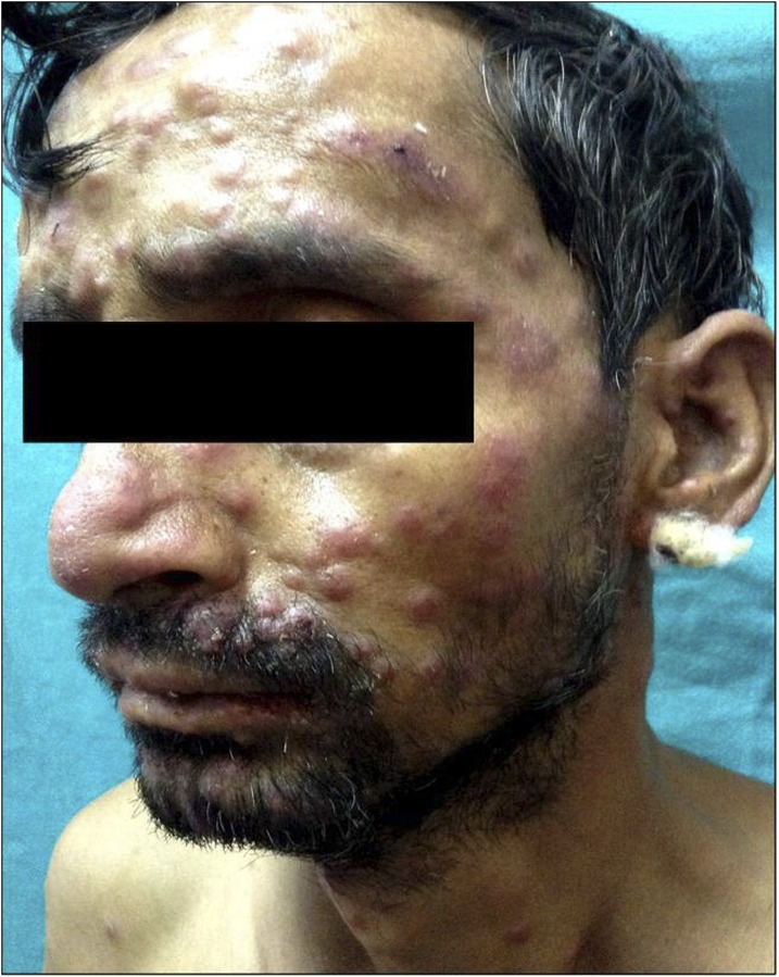
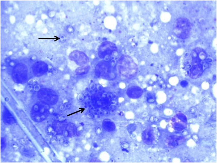
We describe four cases of histoplasmosis indigenous to Himachal Pradesh (India) that will be of considerable public health interest. A 48-year-old human immunodeficiency virus (HIV)-negative man with cervical and mediastinal lymphadenopathy, hepatosplenomegaly, adrenal mass, and bone marrow involvement was treated as disseminated tuberculosis without benefit. Progressive disseminated histoplasmosis was diagnosed from the fungus in smears from adrenal mass. Another 37-year-old HIV-positive man was on treatment of suspected pulmonary tuberculosis. He developed numerous erythema nodosum leprosum-like mucocutanous lesions accompanied by fever, generalized lymphadenopathy, and weight loss. Pulmonary histoplasmosis with cutaneous dissemination was diagnosed when skin lesions showed the fungus in smears, histopathology, and mycologic culture. Both were successfully treated with amphotericin B/itraconazole. Third patient, a 46-year-old HIV-negative man, had oropharyngeal lesions, cervical lymphadenopathy, intermittent fever, hepatosplenomegaly, and deteriorating general health. Progressive disseminated oropharyngeal histoplasmosis was diagnosed from the fungus in smears and mycologic cultures from oropharyngeal lesions and cervical lymph node aspirates. He died despite initiating treatment with oral itraconazole. Another 32-year-old man 3 months after roadside trauma developed a large ulcer with exuberant granulation tissue over left thigh without evidence of immunosuppression/systemic involvement. He was treated successfully with surgical excision of ulcer under amphotericin B/itraconazole coverage as primary cutaneous histoplasmosis confirmed pathologically and mycologically. A clinical suspicion remains paramount for early diagnosis of histoplasmosis particularly in a nonendemic area. Most importantly, with such diverse clinical presentation and therapeutic outcome selection of an appropriate and customized treatment schedule is a discretion the treating clinicians need to make.
Figures


References
-
- Kasuga T, et al. 2003. Phylogeography of the fungal pathogen Histoplasma capsulatum. Mol Ecol 12: 3383–3401. - PubMed
-
- Fujio J, Nishimura K, Miyaji M, 1999. Epidemiological survey of the imported mycoses in Japan. Jpn J Med Mycol 40: 103–109. - PubMed
-
- Kathuria S, Capoor MR, Yadav S, Singh A, Ramesh V, 2013. Disseminated histoplasmosis in an apparently immunocompetent individual from north India: a case report and review. Med Mycol 51: 774–778. - PubMed
-
- Randhawa HS, Khan ZU, 1994. Histoplasmosis in India: current status. Indian J Chest Allied Sci 36: 193–213. - PubMed
Publication types
MeSH terms
Substances
LinkOut - more resources
Full Text Sources
Other Literature Sources
Medical
