

Nigrosome 1 visibility at susceptibility weighted 7T MRI-A dependable diagnostic marker for Parkinson's disease or merely an inconsistent, age-dependent imaging finding?
- PMID: 29016618
- PMCID: PMC5634553
- DOI: 10.1371/journal.pone.0185489
Nigrosome 1 visibility at susceptibility weighted 7T MRI-A dependable diagnostic marker for Parkinson's disease or merely an inconsistent, age-dependent imaging finding?
Abstract
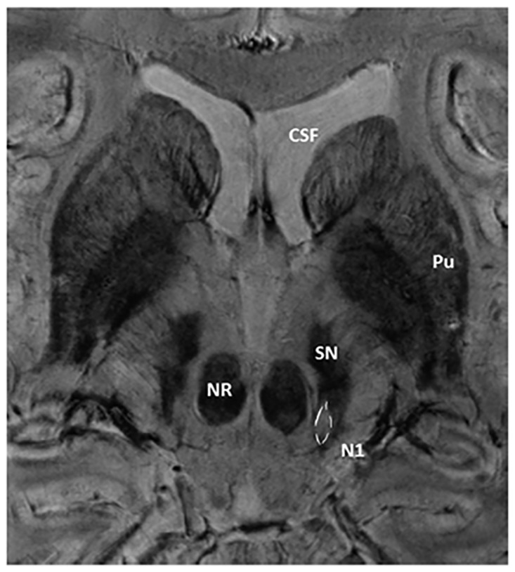
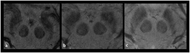
Background: Visualisation of nigrosome 1, a substructure of the healthy substantia nigra, was restricted in susceptibility weighted MR imaging in almost all patients with Parkinson's disease studied so far. The purpose of this study was to determine the degree of visibility of this substructure in subjects without Parkinson's disease and to examine the potential link between increasing brain iron accumulation with age and its detectability.
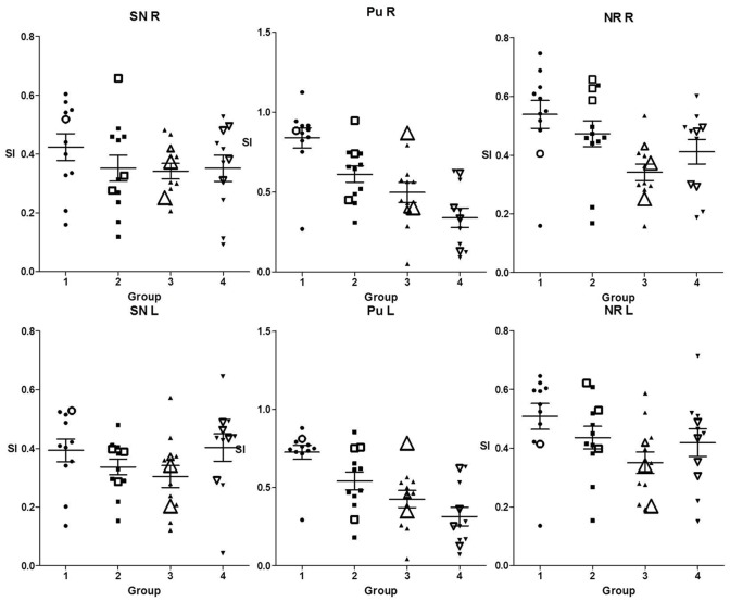
Methods: In 46 subjects (21 women, 25 men; 19 to 75 y; mean age: 44.5; SD: 15.6) examined with susceptibility weighted MR imaging at 7T visibility of nigrosome 1 was rated and classified. We assessed differences related to age and to signal intensities in the substantia nigra, red nucleus and putamen as correlates of the individual iron concentration.
Results: In 93% nigrosome 1was at least unilaterally clearly present. In 24% at least one-sided limited visibility was observed. Using predefined classification criteria the specificity of the visibility across all age groups reached approximately 94%. We found no correlation with increasing iron concentrations with age.
Conclusion: Aging with a related increase in iron concentration probably does not affect the visibility of nigrosome 1 at 7T SWI MRI. Our results support the role of this feature as a future differential diagnostic tool but further large-scale prospective studies are needed to better define the extent of a "limited visibility" to which an individual can be considered healthy.
Conflict of interest statement
Figures

Similar articles
-
Imaging of nigrosome 1 in substantia nigra at 3T using multiecho susceptibility map-weighted imaging (SMWI).J Magn Reson Imaging. 2017 Aug;46(2):528-536. doi: 10.1002/jmri.25553. Epub 2016 Nov 11. J Magn Reson Imaging. 2017. PMID: 27859983
-
Parkinson's disease related signal change in the nigrosomes 1-5 and the substantia nigra using T2* weighted 7T MRI.Neuroimage Clin. 2018 May 24;19:683-689. doi: 10.1016/j.nicl.2018.05.027. eCollection 2018. Neuroimage Clin. 2018. PMID: 29872633 Free PMC article.
-
A preliminary attempt to visualize nigrosome 1 in the substantia nigra for Parkinson's disease at 3T: An efficient susceptibility map-weighted imaging (SMWI) with quantitative susceptibility mapping using deep neural network (QSMnet).Med Phys. 2020 Mar;47(3):1151-1160. doi: 10.1002/mp.13999. Epub 2020 Jan 30. Med Phys. 2020. PMID: 31883389
-
Nigrosome 1 imaging: technical considerations and clinical applications.Br J Radiol. 2019 Sep;92(1101):20180842. doi: 10.1259/bjr.20180842. Epub 2019 Jun 5. Br J Radiol. 2019. PMID: 31067082 Free PMC article. Review.
-
7 Tesla magnetic resonance imaging: a closer look at substantia nigra anatomy in Parkinson's disease.Mov Disord. 2014 Nov;29(13):1574-81. doi: 10.1002/mds.26043. Epub 2014 Oct 12. Mov Disord. 2014. PMID: 25308960 Review.
Cited by
-
The clinical application of nigrosome 1 detection on high-resolution susceptibility-weighted imaging in the evaluation of suspected Parkinsonism: The real-world performance and pitfalls.PLoS One. 2020 Apr 2;15(4):e0231010. doi: 10.1371/journal.pone.0231010. eCollection 2020. PLoS One. 2020. PMID: 32240236 Free PMC article.
-
Studying Alzheimer disease, Parkinson disease, and amyotrophic lateral sclerosis with 7-T magnetic resonance.Eur Radiol Exp. 2021 Aug 26;5(1):36. doi: 10.1186/s41747-021-00221-5. Eur Radiol Exp. 2021. PMID: 34435242 Free PMC article. Review.
-
Is the Swallow Tail Sign a Useful Imaging Biomarker in Clinical Neurology? A Systematic Review.Mov Disord Clin Pract. 2025 Feb;12(2):134-147. doi: 10.1002/mdc3.14304. Epub 2024 Dec 17. Mov Disord Clin Pract. 2025. PMID: 39688317 Free PMC article.
-
Present and Future of Ultra-High Field MRI in Neurodegenerative Disorders.Curr Neurol Neurosci Rep. 2018 Apr 20;18(6):31. doi: 10.1007/s11910-018-0841-7. Curr Neurol Neurosci Rep. 2018. PMID: 29679161
-
Iron- and Neuromelanin-Weighted Neuroimaging to Study Mitochondrial Dysfunction in Patients with Parkinson's Disease.Int J Mol Sci. 2022 Nov 8;23(22):13678. doi: 10.3390/ijms232213678. Int J Mol Sci. 2022. PMID: 36430157 Free PMC article. Review.
References
-
- Jankovic J, Rajput AH, McDermott MP, Perl DP. The evolution of diagnosis in early Parkinson disease. Parkinson Study Group. Arch Neurol. 2000; 57[3]:369–72. - PubMed
-
- Mahlknecht P, Hotter A, Hussl A, Esterhammer R, Schocke M, Seppi K. Significance of MRI in diagnosis and differential diagnosis of Parkinson's disease. Neurodegener Dis. 2010; 7[5]:300–18. doi: 10.1159/000314495 - DOI - PubMed
-
- Tolosa E, Wenning G, Poewe W. The diagnosis of Parkinson's disease. Lancet Neurol. 2006; 5[1]:75–86. doi: 10.1016/S1474-4422(05)70285-4 - DOI - PubMed
-
- Seppi K, Yekhlef F, Diem A, Luginger WE, Mueller J, Tison F et al. Progression of parkinsonism in multiple system atrophy. J Neurol. 2005; 252[1]:91–6. doi: 10.1007/s00415-005-0617-2 - DOI - PubMed
MeSH terms
Substances
LinkOut - more resources
Full Text Sources
Other Literature Sources
Medical
