. 2018 Jan 1;114(1):77-89.
doi: 10.1093/cvr/cvx152.

Investigation of cardiac fibroblasts using myocardial slices

Affiliations

Investigation of cardiac fibroblasts using myocardial slices

Filippo Perbellini et al. Cardiovasc Res. .

Abstract

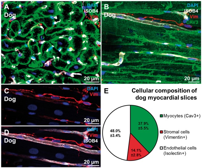
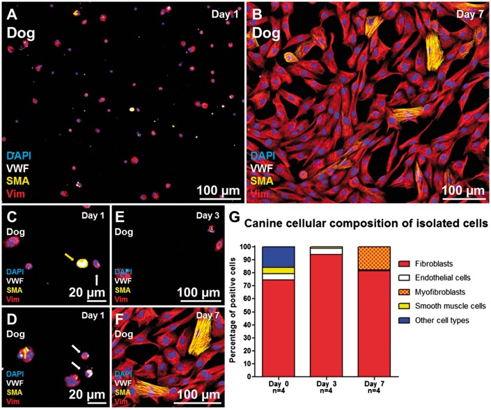
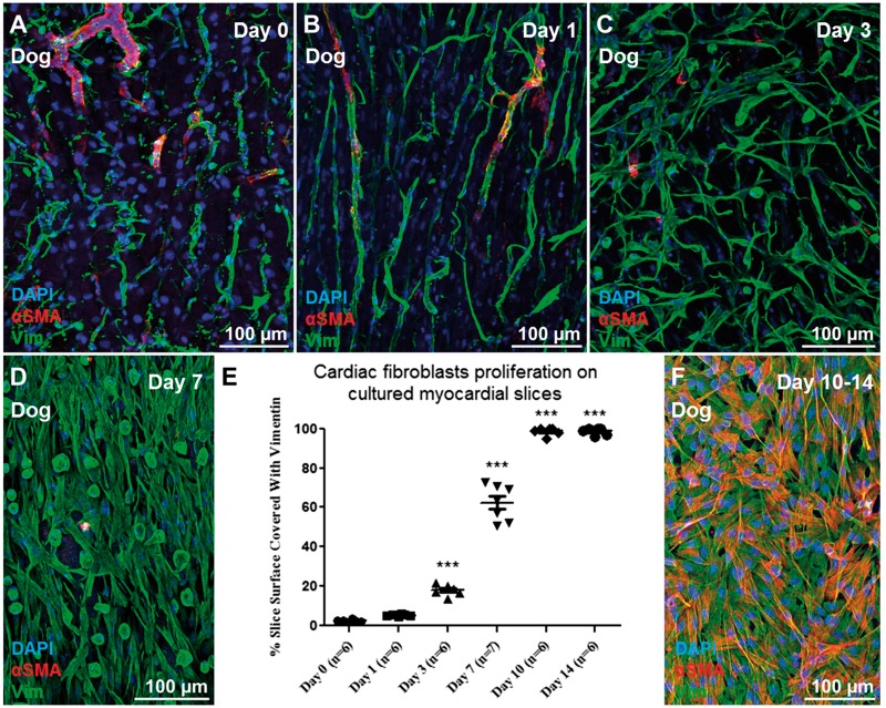
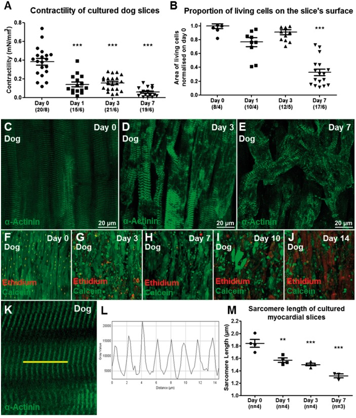
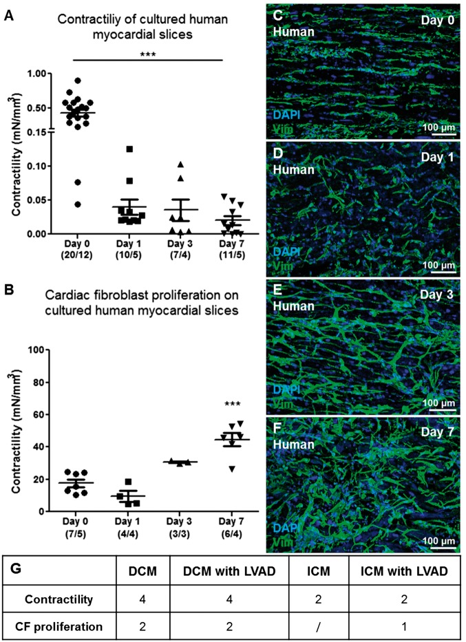
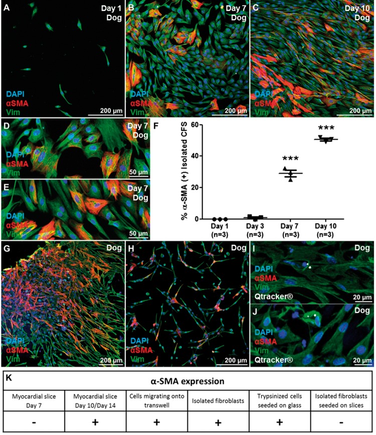
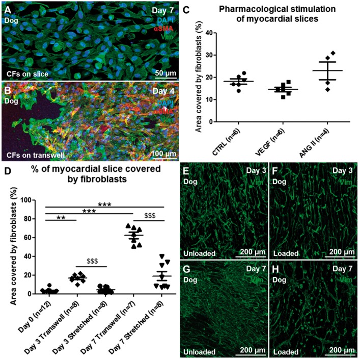
Aims: Cardiac fibroblasts (CFs) are considered the principal regulators of cardiac fibrosis. Factors that influence CF activity are difficult to determine. When isolated and cultured in vitro, CFs undergo rapid phenotypic changes including increased expression of α-SMA. Here we describe a new model to study CFs and their response to pharmacological and mechanical stimuli using in vitro cultured mouse, dog and human myocardial slices.

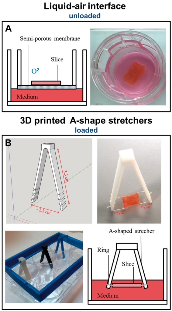
Methods and results: Unloading of myocardial slices induced CF proliferation without α-SMA expression up to 7 days in culture. CFs migrating onto the culture plastic support or cultured on glass expressed αSMA within 3 days. The cells on the slice remained αSMA(-) despite transforming growth factor-β (20 ng/ml) or angiotensin II (200 µM) stimulation. When diastolic load was applied to myocardial slices using A-shaped stretchers, CF proliferation was significantly prevented at Days 3 and 7 (P < 0.001).

Conclusions: Myocardial slices allow the study of CFs in a multicellular environment and may be used to effectively study mechanisms of cardiac fibrosis and potential targets.

Keywords: Cardiac fibroblasts; Fibrosis; Mechanical load; Myocardial slices; α-SMA.


Figures

Figure 1
Figure 1
Cellular composition of dog myocardial slices. (A,B) transverse and longitudinal section of a myocardial slice; cardiomyocytes, endothelial cells and fibroblasts were stained for CAV3 (green), ISOB4 (white), and VIM (red). (C,D) Higher magnification of CFs (red) and their location in proximity to capillaries (white). (E) The cardiac cellular composition is made up by 48% endothelial cells, 14.2% stromal cells, 37.9% myocytes. 44 images generated from 10 myocardial slices were processed and a total of 3259 cells were counted.
Figure 2
Figure 2
Cellular composition of isolated cells after enzymatic digestion of myocardial tissue. (A,B) Isolated cells at Days 1 and 7 stained for VIM (red), αSMA (yellow), and VWF (white). (C–F) higher magnification of isolated cells, the yellow arrow indicates a smooth muscle cell (SMA+), the white arrows indicate endothelial cells (VWF+) (G) after enzymatic digestion 74.5% of the isolated cells were CFs (VIM+/αSMA−/VWF−), 5% were smooth muscle cells (VIM−/αSMA+/VWF−) and 4.5% were endothelial cells (VIM−/αSMA−/VWF+). The remaining 16% of the cells were negative for all the antibodies (VIM−/αSMA−/VWF−) and included cardiomyocytes and other cell types. After 10 days in culture almost exclusively VIM+ cells survive and the cells start to express αSMA (yellow). Two bottom glass dishes were prepared for each time point of each isolation (four isolations) and averaged before statistical analysis.
Figure 3
Figure 3
CFs proliferation on myocardial slices. Representative images of (A–D) VIM+ cell (green) proliferation on the myocardial slice with time. (E) We observed a significant increase in the percentage of the slice surface covered by VIM+ cells at Day 3, with a further significant increase seen at Days 7 and 10. (F) By Day 10 the cells completely covered the surface of the slices and started to express αSMA (red). Three images per slice were acquired and the data obtained were averaged before statistical analysis. Six slices were used for this experiment.
Figure 4
Figure 4
Characterization of myocardial slices. The slices responded to prolonged in vitro culture under unloading conditions (A) with significantly decreased contractility (36%) for the first 3 days and progressively deteriorated to 15% by Day 7. Number of slices/number of experiments. (B, F–J) The viability of the slice was preserved until Day 7, when a significant decrease in the area of living cells was measured (three images per slice were acquired and the data obtained were averaged before statistical analysis. Number of slices/number of experiments; P < 0.001). Immunofluorescence staining for sarcomeric α-actinin (Green) showed myocytes size changes and sarcomeric reorganization occurs with time, suggesting cardiomyocytes dedifferentiation (C–E). The sarcomere length of myocardial slices showed a significant decrease within 24 h in culture (9 K–M). Three images per slice were acquired and the sarcomere length of at least five cells per image was measured, the data were averaged before statistical analysis.
Figure 5
Figure 5
CFs proliferation and contractile capacity of culture human myocardial slices. Human myocardial slices cultured in vitro progressively lose their contractility (A). Number of slices/number of experiments. As observed in canine myocardial slices, we noticed a significant increase in the percentage of the slice surface covered by VIM+ cells (B). Representative images of VIM+ cells proliferating on the human slice surface at Days 0, 1, 3, and 7 (Green, C–F). Three images were acquired from each slice and the data obtained were averaged before statistical analysis. The slices were prepared from at least three human samples (K). The Table summarizes the groups of end-stage heart patient biopsies used in the study. (G) DCM, dilated cardiomyopathy; DCM with LVAD, dilated cardiomyopathy with left ventricular assist device; ICM, ischaemic cardiomyopathy; ICM with LVAD: ischaemic cardiomyopathy with left ventricular assist device.
Figure 6
Figure 6
Isolated CFs in vitro. Freshly isolated dog CFs (VIM+/αSMA−), were seeded on bottom glass dishes coated with fibronectin. (A–E) Within 3 days, CFs started to express αSMA. By Day 7 30% of the cells were αSMA+ and the number increased to 50% by Day 10 (F). The experiment was repeated three times and three images were acquired from each dish and the data obtained were averaged before statistical analysis. VIM+ cells that proliferated out of the myocardial slices and onto the Transwells membrane expressed αSMA at Day 7. (G). VIM+ cells, trypsinized from Day 7 slice surface and cultured on glass, expressed αSMA after 24 h (H). The cells were isolated after collagenase digestion, labelled with Qtracker 585 (white) and seeded onto dog slices (I, J). Labelled CFs were then cultured for 7 days and observed to be αSMA−. (K) CF αSMA expression was assessed in different conditions. CF proliferating on the slice surface were αSMA(−) after 7 days of in vitro culture. CFs migrating onto the culture plastic support or isolated with enzymatic digestion and cultured on glass were αSMA(+). CFs proliferating on myocardial slices for 7 days [αSMA(−)] were trypsinized and seeded onto glass dishes and on myocardial slices. These cells were respectively αSMA(+) and αSMA(−) suggesting a crucial role in the slice as a substrate to preserve a more physiological phenotype. Each experiment was repeated at least three times.
Figure 7
Figure 7
Graphical and photographic representation of myocardial slices culture method. Transwells membrane (A) were used for the liquid–air interface culture method. With this culture condition the slices were mechanically unloaded. (B) 3D printed A-shape stretchers. These devices were used to apply mechanical load to myocardial slices. PTFE-coated silver rings holders were attached to the edges of each slice, perpendicular to myofibril direction. The lateral notches allow a progressive stretch of the slice.
Figure 8
Figure 8
Pharmacological and mechanical stimulation of myocaridial slices. (A) The pharmacological stimulation of myocardial slices with pro-fibrotic drugs at concentrations that induce myofibroblast phenotype in isolated CFs did not induce αSMA expression in the proliferating CFs. (B) VIM+ cells that proliferate out of the myocardial slice (VIM+ cells, green) and onto the Transwells membrane express αSMA (red). (D) Mechanical stretch of myocardial slices induced a significant reduction in fibroblast proliferation at both Days 3 and 7. Representative images of CFs stained for VIM (green) proliferating on the surface of myocardial slices at Days 3 (E unloaded, F loaded) and 7 (G unloaded, H loaded). Each experiment was repeated at least three times, to quantify CF proliferation three images per slice were acquired and the data obtained were averaged before statistical analysis.

References

    1. Biernacka A, Frangogiannis NG.. Aging and Cardiac Fibrosis. Aging Dis 2011;2:158–173. - PMC - PubMed
    1. Upadhya B, Taffet GE, Cheng CP, Kitzman DW.. Heart failure with preserved ejection fraction in the elderly: scope of the problem. J Mol Cell Cardiol 2015;83:73–87. - PMC - PubMed
    1. Abramochkin DV, Lozinsky IT, Kamkin A.. Influence of mechanical stress on fibroblast-myocyte interactions in mammalian heart. J Mol Cell Cardiol 2014;70:27–36. - PubMed
    1. Cartledge J. The Effects of Cardiac Fibroblasts on Cardiac Myocyte Structure and Excitation-Contraction Coupling Through Paracrine Mediators. National Heart and Lung Institute; 2013.
    1. Shinde AV, Frangogiannis NG.. Fibroblasts in myocardial infarction: a role in inflammation and repair. J Mol Cell Cardiol 2014;70:74–82. - PMC - PubMed

Publication types

MeSH terms