. 2017 Oct 15;26(20):3935-3945.
doi: 10.1093/hmg/ddx281.

Potassium channel dysfunction underlies Purkinje neuron spiking abnormalities in spinocerebellar ataxia type 2

Affiliations

Potassium channel dysfunction underlies Purkinje neuron spiking abnormalities in spinocerebellar ataxia type 2

James M Dell'Orco et al. Hum Mol Genet. .

Abstract

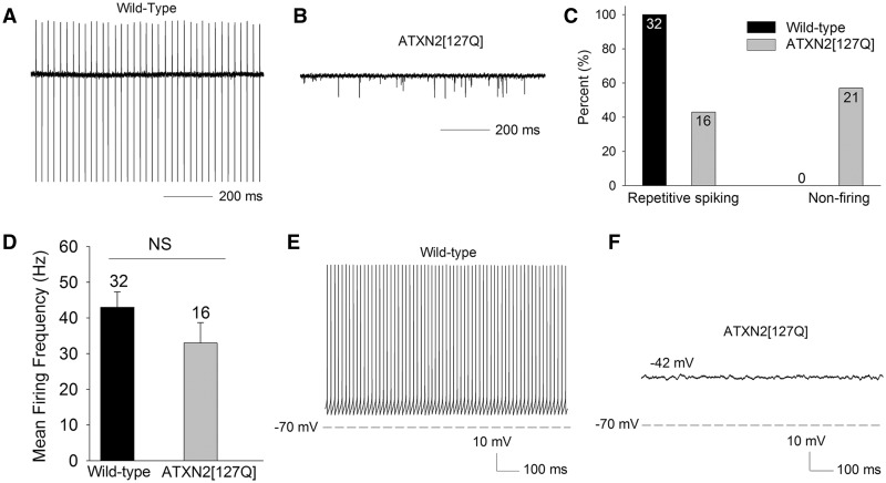
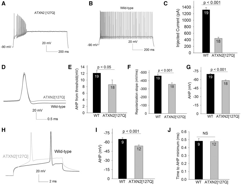
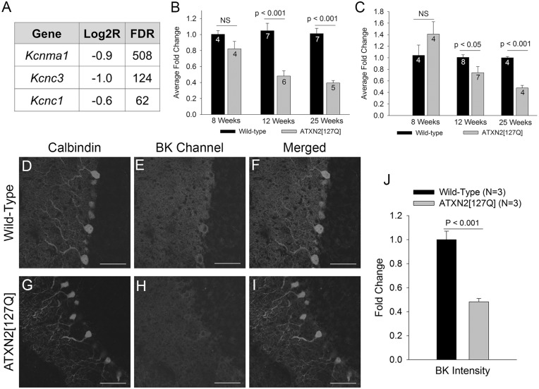
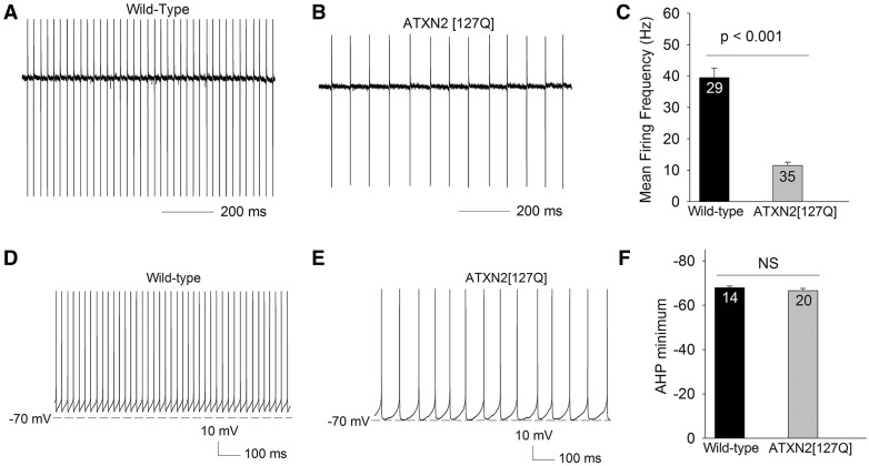
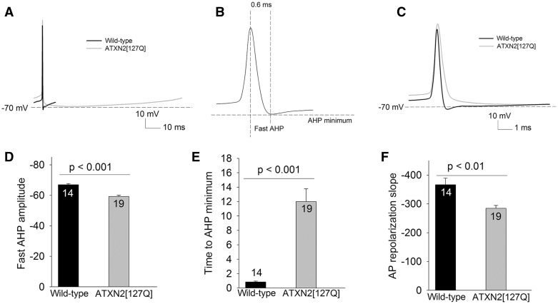
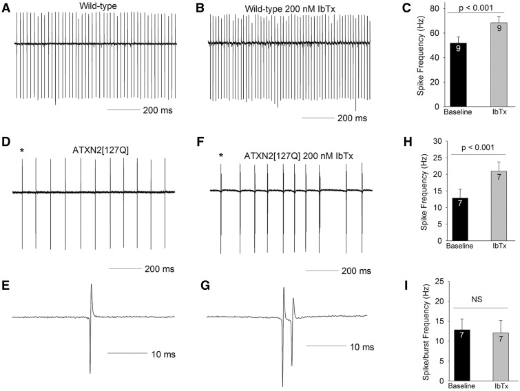
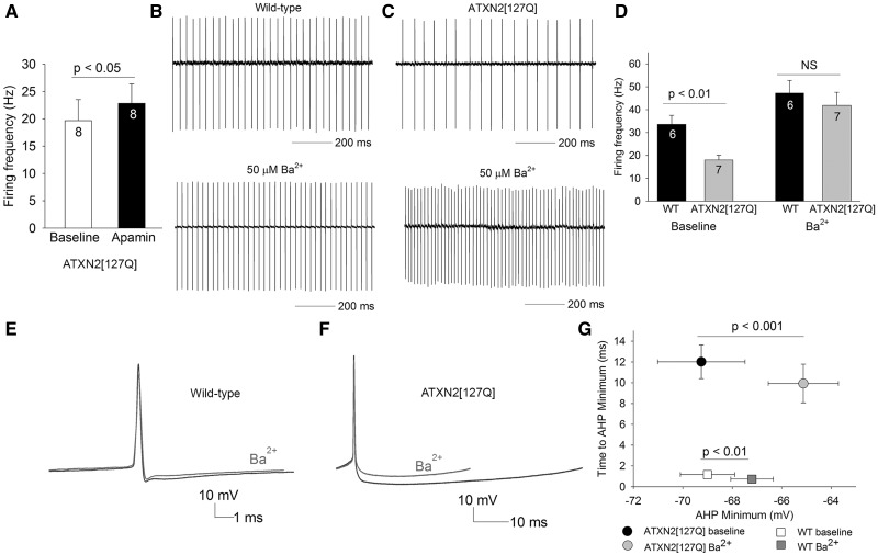
Alterations in Purkinje neuron firing often accompany ataxia, but the molecular basis for these changes is poorly understood. In a mouse model of spinocerebellar ataxia type 2 (SCA2), a progressive reduction in Purkinje neuron firing frequency accompanies cell atrophy. We investigated the basis for altered Purkinje neuron firing in SCA2. A reduction in the expression of large-conductance calcium-activated potassium (BK) channels and Kv3.3 voltage-gated potassium channels accompanies the inability of Purkinje neurons early in disease to maintain repetitive spiking. In association with prominent Purkinje neuron atrophy, repetitive spiking is restored, although at a greatly reduced firing frequency. In spite of a continued impairment in spike repolarization and a persistently reduced BK channel mediated afterhyperpolarization (AHP), repetitive spiking is maintained, through the increased activity of barium-sensitive potassium channels, most consistent with inwardly rectifying potassium (Kir) channels. Increased activity of Kir channels results in the generation of a novel AHP not seen in wild-type Purkinje neurons that also accounts for the reduced firing frequency late in disease. Homeostatic changes in Purkinje neuron morphology that help to preserve repetitive spiking can also therefore have deleterious consequences for spike frequency. These results suggest that the basis for spiking abnormalities in SCA2 differ depending on disease stage, and interventions targeted towards correcting potassium channel dysfunction in ataxia need to be tailored to the specific stage in the degenerative process.


Figures

Figure 1
Figure 1
ATXN2[127Q] Purkinje neurons exhibit impaired spiking at 12 weeks. (A) Wild-type Purkinje neurons exhibit repetitive spiking (B) A significant proportion of ATXN2[127Q] Purkinje neurons lack spiking at 12 weeks, summarized in (C). (D) The firing frequency of firing ATXN2[127Q] Purkinje neurons is not statistically significantly reduced at this time point. (E) In the whole-cell configuration wild-type Purkinje neurons continue to display regular, repetitive spiking. (F) Non-firing ATXN2[127Q] Purkinje neurons display a depolarized membrane potential. Data were analysed with a Student’s t-test, and are displayed as mean ± SEM.
Figure 2
Figure 2
Impaired spiking in 12-week ATXN2[127Q] Purkinje neurons is secondary to loss of repolarizing potassium conductances. (A) In response to depolarizing current injection of 650 pA from a negative holding potential of − 90 mV, ATXN2[127Q] Purkinje neurons can generate spikes, but cannot sustain spike trains. (B) Purkinje neurons from wild-type littermate controls can sustain high rates of repetitive firing in response to the same amount of depolarizing current injection. (C) The amount of injected current needed for ATXN2[127Q] Purkinje neurons to undergo depolarization block of repetitive spiking is significantly lower than in wild-type littermate controls. (D) The inability of ATXN2[127Q] Purkinje neurons to sustain spike trains is associated with slower spike repolarization and a reduction the amplitude of the AHP. For clarity, traces with similar spike threshold in ATXN2[127Q] and wild-type Purkinje neurons are shown. (E) Summary of AHP amplitude from threshold. (F) Summary of spike repolarization slope. (G) The absolute value of the AHP is more depolarized in ATXN2[127Q] Purkinje neurons. (H) Single spikes were evoked using 10 ms depolarizing steps from −90 mV. Note the reduction in AHP amplitude in ATXN2[127Q] Purkinje neurons, without a change in spike height or the time to minimum AHP, summarized in (I, J) Numbers within the bars indicate numbers of cells. Data were analysed with a Student’s t-test, and are displayed as mean ± SEM.
Figure 3
Figure 3
A progressive reduction in potassium channel transcripts accompanies degeneration in ATXN2[127Q] Purkinje neurons. (A) RNA sequencing from 6-week-old cerebella of ATXN2[127Q] mice and littermate controls reveals a reduction in transcripts of potassium channels important for Purkinje neuron spiking. FDR (false discovery rate) >30 corresponds to a corrected p value of < 0.01. (B) Quantitative RT-PCR for Kcnma1 (BK) demonstrates a progressive reduction in BK channel transcripts in ATXN2[127Q] cerebella. (C) Quantitative RT-PCR for Kcnc3 (Kv3.3) demonstrates a progressive reduction in Kv3.3 channel transcripts in ATXN2[127Q] cerebella. Immunostaining for calbindin (D) and BK channels (E) in wild-type Purkinje neurons shows prominent overlap of BK and calbindin staining (F). (G) In 25-week-old ATXN2[127Q] cerebella, calbindin immunostaining reveals prominent Purkinje neuron dendritic atrophy, with thinning of the molecular layer. (H) BK staining is reduced in ATXN2[127Q] Purkinje neurons, also seen in the merged image of BK and calbindin in (I) and summarized in (J). Data were analysed with a Student’s t-test, and are displayed as mean ± SEM.
Figure 4
Figure 4
Atrophic 25-week ATXN2[127Q] Purkinje neurons are able to restore spiking in association with a normal AHP amplitude. (A) In the cell-attached configuration, wild-type Purkinje neurons at 25 weeks continue to display tonic repetitive spiking. (B) ATXN2[127Q] Purkinje neurons also display tonic repetitive spiking at 25 weeks, although at a greatly reduced firing frequency, summarized in (C). (D) In the whole-cell configuration wild-type Purkinje neurons display spikes with a prominent AHP. (E) ATXN2[127Q] Purkinje neurons display repetitive spiking with a similar amplitude AHP, summarized in (F). Data were analysed with a Student’s t-test, and are displayed as mean ± SEM.
Figure 5
Figure 5
25-week ATXN2[127Q] Purkinje neurons generate an AHP with novel kinetics. (A) Overlay of one interspike interval from a wild-type and ATXN2[127Q] Purkinje neuron showing a similar AHP amplitude. (B) Schematic action potential showing the location of the fast AHP and the AHP minimum. (C) Overlay of a spontaneous spike from a wild-type and ATXN2[127Q] Purkinje neuron on an expanded timescale to demonstrate the slowing of spike repolarization and reduced fast AHP amplitude in ATXN2[127Q] Purkinje neurons. (D) Summary of the fast AHP amplitude in ATXN2[127Q] Purkinje neurons. (E) The AHP minimum is delayed in ATXN2[127Q] Purkinje neurons. (F) The repolarization of the action potential (AP) is impaired in ATXN2[127Q] Purkinje neurons. Data were analysed with a Student’s t-test, and are displayed as mean ± SEM.
Figure 6
Figure 6
BK channels are not responsible for the slow firing in 25-week ATXN2[127Q] Purkinje neurons. (A) In the cell-attached configuration, wild-type Purkinje neurons at 25 weeks continue to display tonic repetitive spiking. (B) In this cell 200 nM iberiotoxin (IbTx) increases firing frequency, summarized in (C). (D) ATXN2[127Q] Purkinje neurons display tonic repetitive spiking at 25 weeks at a reduced firing frequency. (E) A single spike corresponding to the asterisk is shown on an expanded timescale. (F) In this cell, 200 nM iberiotoxin converts tonic spiking into tonic bursting, with each burst containing a spike followed by one or two spikelets. (G) A single spike-burst corresponding to the asterisk is shown on an expanded timescale, showing a spike and a spikelet. (H) Iberiotoxin increases firing frequency by converting spikes into spike-bursts. (I) Burst frequency is similar to the firing frequency of tonic repetitive spiking in the absence of iberiotoxin. Data were analysed with a Student’s t-test, and are displayed as mean ± SEM.
Figure 7
Figure 7
The novel AHP generated by Kir channels is responsible for the reduced firing frequency of 25-week ATXN2[127Q] Purkinje neurons. (A) Apamin (100 nM) has a minimal effect on firing frequency of ATXN2[127Q] Purkinje neurons. (B) Wild-type neurons that display tonic repetitive spiking (top) have a modest increase in firing frequency in the presence of 50 µM extracellular barium. (C) ATXN2[127Q] Purkinje neurons (top) increase their firing frequency to wild-type levels in the presence of 50 µM extracellular barium, summarized in (D). (E) Barium has a modest effect on the AHP in wild-type Purkinje neurons (n = 6). (F) The barium sensitive current contributes the entire AHP in ATXN2[127Q] Purkinje neurons (n = 8), summarized in G. Data were analysed with a Student’s t-test, and are displayed as mean ± SEM.

References

    1. Raman I.M., Bean B.P. (1999) Ionic currents underlying spontaneous action potentials in isolated cerebellar Purkinje neurons. J. Neurosci., 19, 1663–1674. - PMC - PubMed
    1. Hausser M., Clark B.A. (1997) Tonic synaptic inhibition modulates neuronal output pattern and spatiotemporal synaptic integration. Neuron, 19, 665–678. - PubMed
    1. Bell C.C., Grimm R.J. (1969) Discharge properties of Purkinje cells recorded on single and double microelectrodes. J. Neurophysiol., 32, 1044–1055. - PubMed
    1. Thach W.T. (1968) Discharge of Purkinje and cerebellar nuclear neurons during rapidly alternating arm movements in the monkey. J. Neurophysiol., 31, 785–797. - PubMed
    1. Thach W.T. (1978) Correlation of neural discharge with pattern and force of muscular activity, joint position, and direction of intended next movement in motor cortex and cerebellum. J. Neurophysiol., 41, 654–676. - PubMed

Publication types