. 2017 Oct 9;43(1):48-59.e5.
doi: 10.1016/j.devcel.2017.09.003.

Fgf10-Hippo Epithelial-Mesenchymal Crosstalk Maintains and Recruits Lung Basal Stem Cells

Affiliations

Fgf10-Hippo Epithelial-Mesenchymal Crosstalk Maintains and Recruits Lung Basal Stem Cells

Thomas Volckaert et al. Dev Cell. .

Abstract

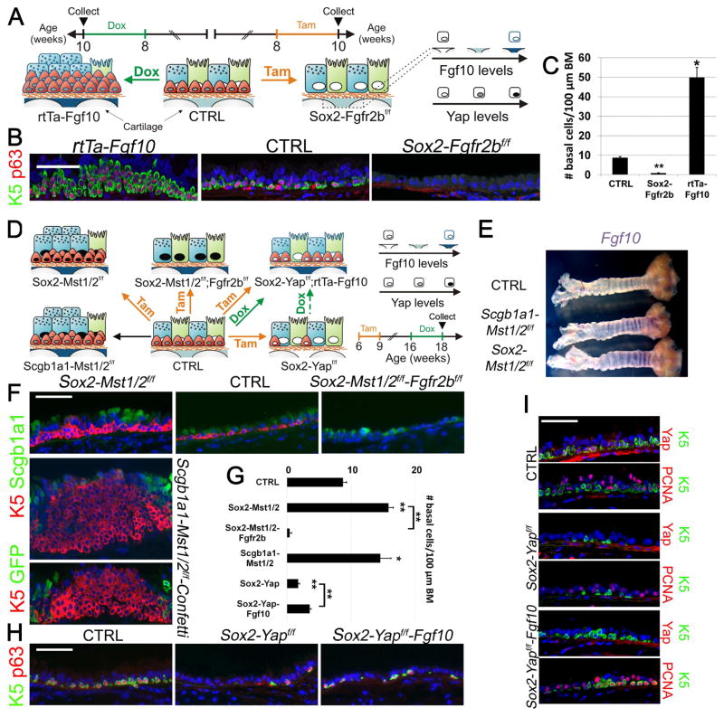
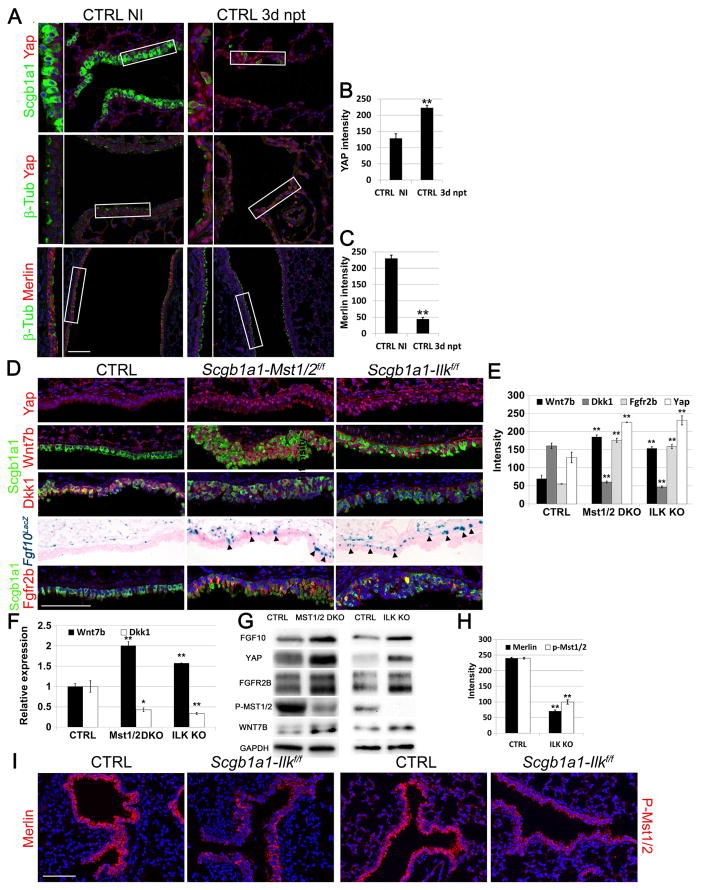
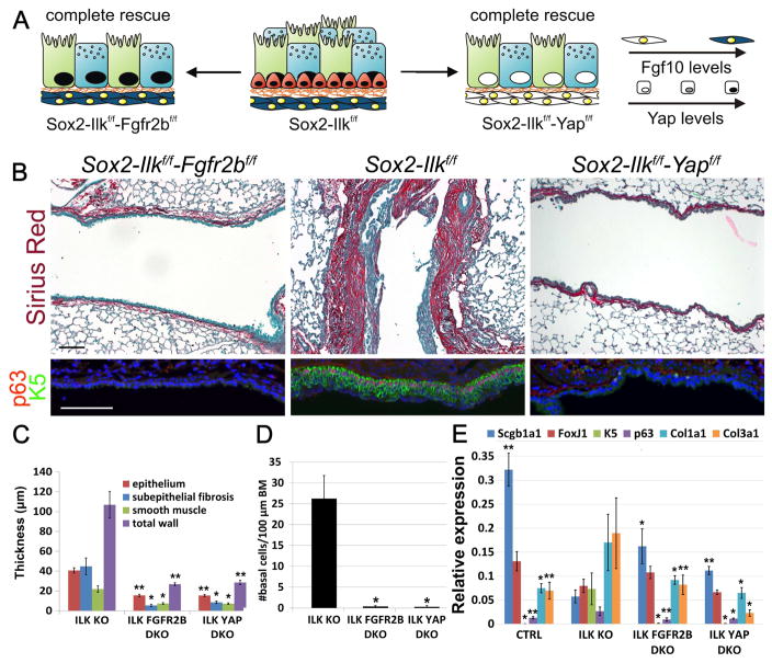
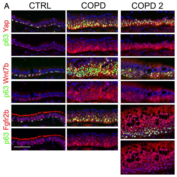
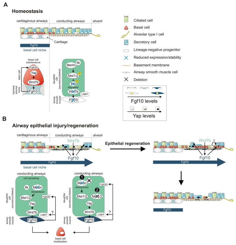
The lung harbors its basal stem/progenitor cells (BSCs) in the protected environment of the cartilaginous airways. After major lung injuries, BSCs are activated and recruited to sites of injury. Here, we show that during homeostasis, BSCs in cartilaginous airways maintain their stem cell state by downregulating the Hippo pathway (resulting in increased nuclear Yap), which generates a localized Fgf10-expressing stromal niche; in contrast, differentiated epithelial cells in non-cartilaginous airways maintain quiescence by activating the Hippo pathway and inhibiting Fgf10 expression in airway smooth muscle cells (ASMCs). However, upon injury, surviving differentiated epithelial cells spread to maintain barrier function and recruit integrin-linked kinase to adhesion sites, which leads to Merlin degradation, downregulation of the Hippo pathway, nuclear Yap translocation, and expression and secretion of Wnt7b. Epithelial-derived Wnt7b, then in turn, induces Fgf10 expression in ASMCs, which extends the BSC niche to promote regeneration.

Keywords: Fgf10; Fgfr2; Hippo; Ilk; Yap; basal stem cell; integrin; lung; regeneration; stem cell niche.


Conflict of interest statement

The authors declare no conflict of interest

Figures

Figure 1
Figure 1. The inactive Hippo pathway in basal stem/progenitor cells generates the Fgf10-expressing tracheal stromal niche required to maintain their cell pool
(A) Experimental strategy and schematic representation of tracheal BSC amplification after Fgf10 overexpression or tracheal BSC loss after Fgfr2b ablation in airway epithelial cells. Ciliated, secretory and BSCs are shown in green, blue and red, respectively. (B) Immunostaining on rtTa-Fgf10, control and Sox2-Fgfr2bf/f tracheas for the BSC markers Keratin 5 (K5) (green) and p63 (red) 14 days after doxycycline or tamoxifen induction. (C) Quantification of the number (#) of BSCs per 100 μm basement membrane of pictures represented in (B). (D) Experimental strategy and schematic representation of Mst1/2 ablation in all airway epithelial cells or selectively in secretory/ciliated cells alone or in combination with either Fgfr2b or Yap in all airway epithelial cells with or without simultaneously inducing Fgf10 expression. (E) Whole mount in situ hybridization for Fgf10 in control, Scgb1a1-Mst1/2f/f and Sox2-Mst1/2f/f tracheas. Note purple Fgf10 expression between the tracheal cartilage rings. (F) Upper panels show immunostaining on control, Sox2-Mst1/2f/f and Sox2-Mst1/2f/f-Fgfr2bf/f tracheas 2 months after tamoxifen induction as well as on a 2.5 month old Scgb1a1-Mst1/2f/f-Confetti trachea for the BSC marker K5 (red) and the secretory cell marker Scgb1a1 (green). Lower panel shows immunostaining on Scgb1a1-Mst1/2f/f-Confetti trachea for the BSC marker K5 (red) and GFP (green). (G) Quantification of the number (#) of BSCs per 100 μm basement membrane (BM) of pictures represented in (F,H). (H) Immunostaining on control, Sox2-Yapf/f, Sox2-Yapf/f-rtTa-Fgf10 tracheas for the BSC markers K5 (green) and p63 (red) after tamoxifen-induced deletion of Yap and/or doxycycline-induced Fgf10 expression. Refer to panel D for experimental strategy. (I) Immunostaining on adjacent tracheal sections from control, Sox2-Yapf/f and Sox2-Yapf/f-Fgf10 mice for BSC marker K5 (green) and Yap (red) or proliferation marker PCNA (red) 12 weeks after tamoxifen and 2 weeks after doxycycline induction, starting at 10 weeks after tamoxifen induction. Nuclei, DAPI (blue). **P < 0.01; *P < 0.05. n ≥ 6.; error bars mean ± SEM. Scale bars, 100 μm (B,F,H); 50 μm (I). (See also Figure S1)
Figure 2
Figure 2. Inactivation of the Hippo pathway in differentiated airway epithelial cells after injury or after Ilk inactivation induces epithelial Wnt7b expression and Fgf10 secretion by ASMCs
(A) Immunostaining on non-cartilaginous airways of non-injured (NI) control lungs and lungs 3 days after naphthalene (npt)-induced injury for secretory cell marker Scgb1a1 (green) or ciliated cell marker β-tubulin (green) and Yap or Merlin (red). (B) Quantification of Yap pixel intensity of pictures represented in (A) (n ≥ 6 mice). (C) Quantification of Merlin pixel intensity of pictures represented in (A) (n ≥ 6 mice). (D) Immunostaining on non-cartilaginous airways of control Fgf10LacZ, Scgb1a1-Mst1/2f/f-Fgf10LacZ and Scgb1a1-Ilkf/f-Fgf10LacZ lungs for secretory cell marker Scgb1a1 (green) with either Yap (red) or Wnt7b (red) or Dkk1 (red) or Fgfr2b (red) and β-gal staining of Fgf10LacZ. Black arrowheads indicate Fgf10 expression in ASMCs. (E) Quantification of pixel intensity of Yap, Wnt7b, Dkk1 and Fgfr2b signals represented in (D) (n ≥ 6 mice). (F) Relative mRNA expression of Wnt7b and Dkk1 in control, Scgb1a1-Mst1/2f/f and Scgb1a1-Ilkf/f lungs. (G) Western blot analysis showing FGF10, YAP, FGFR2B, P-MST1/2, and WNT7B protein expression in control, Scgb1a1-Mst1/2f/f (MST1/2 KO) and Scgb1a1-Ilkf/f (ILK KO) lungs. (H) Quantification of pixel intensities of pictures represented in (I) (n ≥ 6 mice). (I) Immunostaining on control and Scgb1a1-Ilkf/f non-cartilaginous airways for Merlin (red) or phospho-Mst1/2 (red). Nuclei, DAPI (blue). **P < 0.01; *P < 0.05. n ≥ 6.; error bars mean ± SEM. Scale bar, 100 μm. (See also Figures S2, S3, S4, S6 and S7)
Figure 3
Figure 3. Fgf10 overexpression or Ilk deletion in secretory cells induces basal cell recruitment to the non-cartilaginous conducting airways
(A) Immunostaining for Scgb1a1 (green) and p63 (red) (left), or Scgb1a1 (green) and K5 (red) (middle), or Scgb1a1 (green) and Sftpc (red) (right) on non-cartilaginous airways of Scgb1a1-rtTaf/f-Fgf10 mice, which were induced with doxycycline for 5.5 weeks starting at 3 weeks of age. (A′) High magnification of selected regions in A. (B) Immunostaining on control and Scgb1a1-Ilkf/f non-cartilaginous airways for Scgb1a1 (green) and K5 (red) or K5 (red) by itself. (B′) High magnification of selected regions in B. Nuclei were stained with DAPI (blue). Scale bar, 500 μm (A,C), 100 μm (A′), 300 μm (B′) (See also Figure S5).
Figure 4
Figure 4. Fgf10 expression in ASMCs drives secretory and basal cell amplification in the lower non-cartilaginous conducting airways upon Ilk inactivation
(A) Schematic representation of airway BSC amplification or loss, stromal Fgf10 or nuclear Yap levels in the different mutant strains. (B) Sirius red (collagen)/fast green staining and immunostaining on Scgb1a1-Ilkf/f (ILK KO), Scgb1a1-Ilkf/f-Fgfr2bf/f (ILK FGFR2b DKO) and Scgb1a1-Ilkf/f-rtTa-Dkk1 (ILK DKK1) non-cartilaginous airways for BSC markers p63 (red) and Keratin 5 (K5) (green) or secretory cell marker Scgb1a1 (green) and Yap (red). (C,D) Morphometric analysis of airway remodeling (C) and quantification of BSC numbers (D) of images represented in (B). (E) Relative mRNA levels of Scgb1a1 (secretory cells), FoxJ1 (ciliated cells), K5 (BSCs), p63 (BSCs), Col1a1 (collagen) and Col3a1 (collagen) in control, Scgb1a1-Ilkf/f, Scgb1a1-Ilkf/f-Fgfr2bf/f and Scgb1a1-Ilkf/f-rtTa-Dkk1 lungs. Nuclei were stained with DAPI (blue). **P < 0.01; *P < 0.05. n ≥ 6.; error bars mean ± SEM. Scale bars, 100 μm. (See also Figures S3, S4, S6F and S7)
Figure 5
Figure 5. Fgf10-Fgfr2b signaling downstream of Yap drives secretory and basal cell amplification in the lower non-cartilaginous conducting airways upon Ilk inactivation
(A) Schematic representation of non-cartilaginous airway secretory/ciliated amplification and BSC amplification or loss, stromal Fgf10 or nuclear Yap levels upon airway epithelial Ilk/Fgfr2b ablation, Ilk ablation and Ilk/Yap ablation. All lungs were collected at 2 months after tamoxifen induction. (B) Sirius red (collagen)/fast green staining and immunostaining on Sox2-Ilkf/f-Fgfr2bf/f, Sox2-Ilkf/f, and Sox2-Ilkf/f-Yapf/f non-cartilaginous airways for BSC markers p63 (red) and Keratin 5 (K5) (green). (C,D) Morphometric analysis of airway remodeling (C) and quantification of BSC numbers (#) per 100 μm basement membrane (D) from images represented in (B). (E) Relative mRNA levels of Scgb1a1 (secretory cells), FoxJ1 (ciliated cells), K5 (BSCs), p63 (BSCs), Col1a1 (collagen) and Col3a1 (collagen) in control, Sox2-Ilkf/f (ILK KO), Sox2-Ilkf/f-Fgfr2bf/f (ILK FGFR2b DKO) and Sox2-Ilkf/f-Yapf/f (ILK YAP DKO) lungs. Nuclei were stained with DAPI (blue). **P < 0.01; *P < 0.05. n ≥ 6.; error bars mean ± SEM. Scale bars, 100 μm. (See also Figures S3, S4 and S6C)
Figure 6
Figure 6. The Hippo pathway regulates Fgf10 signaling to induce basal stem/progenitor cell amplification in COPD
(A) Immunostaining on control and COPD distal airways for BSC marker p63 (green) and Yap (red) or Wnt7b (red) or Fgfr2b (red). (n=5). Note staining of cilia for Fgfr2b in control but not in COPD airways indicating a loss of ciliated cells in COPD. (See also Table S1)
Figure 7
Figure 7. Model showing how Yap maintains and generates basal like stem cells by activating the stromal niche during homeostasis and epithelial regeneration
(A) During homeostasis, basal stem/progenitor cells are only found in the trachea, where they maintain themselves by inducing Fgf10 expression in the tracheal stromal niche downstream of Yap. Ilk positively regulates the Hippo pathway in conducting airway epithelial cells to maintain their quiescence by inhibiting Yap-mediated activation of Fgf10 expression in ASMCs. (B) Hippo pathway inactivation in conducting non-cartilaginous airway epithelium in response to injury or by deleting Ilk (4) or Mst1/2 (5) results in nuclear Yap localization, increased epithelial Wnt7b expression and Fgf10 expression in ASMCs, which reciprocally breaks epithelial quiescence. The Fgf10-expressing basal stem cell niche extends into the non-cartilaginous airways allowing for BSC mobilization into the lower conducting airways upon airway epithelial Ilk deletion. Alternatively, Fgf10 released by the activated ASMC niche could drive the differentiation of LNEPs, which are scattered along the lower conducting airways, into the basal stem/progenitor cell lineage.

Similar articles

Cited by

References

    1. Scadden DT, Apte U, Gkretsi V, Bowen WC, Mars WM, Luo JH, Donthamsetty S, Orr A, Monga SP, Wu C, Michalopoulos GK. Enhanced liver regeneration following changes induced by hepatocyte-specific genetic ablation of integrin-linked kinase. Hepatology. 2009;50:844–851. - PMC - PubMed
    1. Auerbach O, Stout AP, Hammond EC, Garfinkel L. Changes in bronchial epithelium in relation to cigarette smoking and in relation to lung cancer. N Engl J Med. 1961;265:253–267. - PubMed
    1. Barkauskas CE, Cronce MJ, Rackley CR, Bowie EJ, Keene DR, Stripp BR, Randell SH, Noble PW, Hogan BL. Type 2 alveolar cells are stem cells in adult lung. J Clin Invest. 2013;123:3025–3036. - PMC - PubMed
    1. Bellusci S, Grindley J, Emoto H, Itoh N, Hogan BL. Fibroblast growth factor 10 (FGF10) and branching morphogenesis in the embryonic mouse lung. Development. 1997;124:4867. - PubMed
    1. Belteki G, Haigh J, Kabacs N, Haigh K, Sison K, Costantini F, Whitsett J, Quaggin SE, Nagy A. Conditional and inducible transgene expression in mice through the combinatorial use of Cre-mediated recombination and tetracycline induction. Nucleic Acids Res. 2005;33:e51. - PMC - PubMed

MeSH terms

Substances

LinkOut - more resources