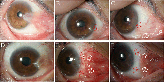
Low recurrence rate of anchored conjunctival rotation flap technique in pterygium surgery
- PMID: 29017515
- PMCID: PMC5634825
- DOI: 10.1186/s12886-017-0587-z
Low recurrence rate of anchored conjunctival rotation flap technique in pterygium surgery
Abstract
Background: To report the recurrence rate for an anchored conjunctival rotation flap technique in primary pterygium surgery.
Methods: Primary pterygium surgeries performed using anchored conjunctival rotation flap techniques (110 eyes in 110 patients) with a minimum follow-up of 12 months were reviewed. In this technique, a conjunctival flap is rotated to cover the bare sclera and suture-fixated with either 8-0 polyglactin (41 eyes) or 10-0 nylon (69 eyes). The recurrence rate was determined, and the two suture materials utilized were compared.
Results: The recurrence rate was 2.71% (3 cases in 110 eyes) when an anchored conjunctival rotation flap technique was used and patients were monitored for 26.40 ± 17.09 months. Interestingly, the recurrences were only observed in polyglactin-sutured eyes. No recurrence was detected in nylon-sutured eyes. No other complications were observed in either group.
Conclusions: The anchored conjunctival rotation flap technique for pterygium surgery has a relatively low recurrence rate. Nylon suture-fixation of the flap was found to be superior to polyglactin suture-fixation in preventing recurrence.
Keywords: Anchored; Flap; Nylon; Polyglactin; Pterygium; Recurrence.
Conflict of interest statement
Ethics approval and consent to participate
This study followed the tenets of the Declaration of Helsinki and was approved by the Institutional Review Board of Dongguk University, Ilsan Hospital, Goyang, South Korea (IRB no. 2016–137).
Consent for publication
Written informed consents were obtained from patients to publish materials and figures included in this study.
Competing interests
The authors declare that they have no competing interests.
Publisher’s Note
Springer Nature remains neutral with regard to jurisdictional claims in published maps and institutional affiliations.
Figures


Similar articles
-
A comparison of anchored conjunctival rotation flap and conjunctival autograft techniques in pterygium surgery.Cornea. 2013 Dec;32(12):1578-81. doi: 10.1097/ICO.0b013e3182a73a48. Cornea. 2013. PMID: 24097183
-
Comparison of autologous fibrin glue versus nylon sutures for securing conjunctival autografting in pterygium surgery.Int Ophthalmol. 2018 Jun;38(3):1219-1224. doi: 10.1007/s10792-017-0585-4. Epub 2017 Jun 17. Int Ophthalmol. 2018. PMID: 28624862 Clinical Trial.
-
Sliding flap of conjunctival limbus to prevent recurrence of pterygium.Refract Corneal Surg. 1992 Sep-Oct;8(5):394-5. Refract Corneal Surg. 1992. PMID: 1450124
-
Options and adjuvants in surgery for pterygium: a report by the American Academy of Ophthalmology.Ophthalmology. 2013 Jan;120(1):201-8. doi: 10.1016/j.ophtha.2012.06.066. Epub 2012 Oct 11. Ophthalmology. 2013. PMID: 23062647 Review.
-
Severe scleral dellen as an early complication of pterygium excision with simple conjunctival closure and review of the literature.Arq Bras Oftalmol. 2014 May-Jun;77(3):182-4. doi: 10.5935/0004-2749.20140046. Arq Bras Oftalmol. 2014. PMID: 25295907 Review.
Cited by
-
Clinical outcome of combined conjunctival autograft transplantation and amniotic membrane transplantation in pterygium surgery.Int J Ophthalmol. 2018 Mar 18;11(3):395-400. doi: 10.18240/ijo.2018.03.08. eCollection 2018. Int J Ophthalmol. 2018. PMID: 29600172 Free PMC article.
-
Pterygium surgery by double-sliding flaps procedure: Comparison between primary and recurrent pterygia.Indian J Ophthalmol. 2021 Sep;69(9):2406-2411. doi: 10.4103/ijo.IJO_2982_20. Indian J Ophthalmol. 2021. PMID: 34427232 Free PMC article.
-
Modified protocol for pterygium surgery without blades and electrocoagulation.Front Med (Lausanne). 2025 Feb 21;12:1522167. doi: 10.3389/fmed.2025.1522167. eCollection 2025. Front Med (Lausanne). 2025. PMID: 40061380 Free PMC article.
-
Pterygium surgery using inferior rotational conjunctival autograft versus conventional conjunctival autograft with sutures - A comparative study.Indian J Ophthalmol. 2023 Dec 1;71(12):3646-3651. doi: 10.4103/IJO.IJO_16_23. Epub 2023 Nov 20. Indian J Ophthalmol. 2023. PMID: 37991298 Free PMC article.
-
Head inversion technique to restore physiological conjunctival structure for surgical treatment of primary pterygium.Sci Rep. 2018 Nov 9;8(1):16603. doi: 10.1038/s41598-018-35121-z. Sci Rep. 2018. PMID: 30413813 Free PMC article.
References
-
- Al Fayez MF. Limbal-conjunctival vs conjunctival autograft transplant for recurrent pterygia: a prospective randomized controlled trial. JAMA Ophthalmol. 2013;131(1):11–6. - PubMed
-
- Hirst LW. Recurrence and complications after 1000 surgeries using pterygium extended removal followed by extended conjunctival transplant. Ophthalmology. 2012;119(11):2205–10. - PubMed
-
- Kheirkhah A, Nazari R, Nikdel M, Ghassemi H, Hashemi H, Behrouz MJ. Postoperative conjunctival inflammation after pterygium surgery with amniotic membrane transplantation versus conjunctival autograft. Am J Ophthalmol. 2011;152(5):733–8. - PubMed
MeSH terms
Substances
LinkOut - more resources
Full Text Sources
Other Literature Sources

