. 2017 Oct 10;7(1):20.
doi: 10.1186/s13395-017-0138-6.

HGF potentiates extracellular matrix-driven migration of human myoblasts: involvement of matrix metalloproteinases and MAPK/ERK pathway

Affiliations

HGF potentiates extracellular matrix-driven migration of human myoblasts: involvement of matrix metalloproteinases and MAPK/ERK pathway

Mariela Natacha González et al. Skelet Muscle. .

Abstract

Background: The hepatocyte growth factor (HGF) is required for the activation of muscle progenitor cells called satellite cells (SC), plays a role in the migration of proliferating SC (myoblasts), and is present as a soluble factor during muscle regeneration, along with extracellular matrix (ECM) molecules. In this study, we aimed at determining whether HGF is able to interact with ECM proteins, particularly laminin 111 and fibronectin, and to modulate human myoblast migration.

Methods: We evaluated the expression of the HGF-receptor c-Met, laminin, and fibronectin receptors by immunoblotting, flow cytometry, or immunofluorescence and used Transwell assays to analyze myoblast migration on laminin 111 and fibronectin in the absence or presence of HGF. Zymography was used to check whether HGF could modulate the production of matrix metalloproteinases by human myoblasts, and the activation of MAPK/ERK pathways was evaluated by immunoblotting.

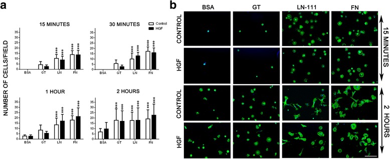
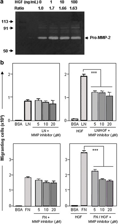
Results: We demonstrated that human myoblasts express c-Met, together with laminin and fibronectin receptors. We observed that human laminin 111 and fibronectin have a chemotactic effect on myoblast migration, and this was synergistically increased when low doses of HGF were added. We detected an increase in MMP-2 activity in myoblasts treated with HGF. Conversely, MMP-2 inhibition decreased the HGF-associated stimulation of cell migration triggered by laminin or fibronectin. HGF treatment also induced in human myoblasts activation of MAPK/ERK pathways, whose specific inhibition decreased the HGF-associated stimulus of cell migration triggered by laminin 111 or fibronectin.

Conclusions: We demonstrate that HGF induces ERK phosphorylation and MMP production, thus stimulating human myoblast migration on ECM molecules. Conceptually, these data state that the mechanisms involved in the migration of human myoblasts comprise both soluble and insoluble moieties. This should be taken into account to optimize the design of therapeutic cell transplantation strategies by improving the migration of donor cells within the host tissue, a main issue regarding this approach.

Keywords: Fibronectin; HGF; Laminin; MAPK/ERK; Matrix metalloproteinases; Migration; Myoblast transplantation.


Conflict of interest statement

Ethics approval and consent to participate

The human myoblasts cells were obtained in accordance with French legislation on ethical rules (authorization AC-2013-1868 given by the bioethics committee of the French ministry).

Consent for publication

Not applicable.

Competing interests

The authors declare that they have no competing interests.

Publisher’s Note

Springer Nature remains neutral with regard to jurisdictional claims in published maps and institutional affiliations.

Figures

Fig. 1
Fig. 1
Phenotypic features of the CHQ human myoblast preparation. a Culture purity was determined by cytofluorometric detection of CD56, a typical myoblast surface marker. As seen in the FACS profile, 87% of this cell preparation did correspond to myoblasts, since labeling was positive for the anti-CD56 antibody, as compared to the black curves, generated after binding of an unrelated antibody. b The myogenicity of the cells was confirmed by desmin expression in immunofluorescence, using an anti-desmin monoclonal antibody. c Immunodetection of c-met by western blotting of whole-cell extracts. Hepatoma cell line HEP-G2: positive control. d Flow cytometry profiles of the expression of CD29 (β1-integrin chain), CD49d, CD49e (integrin α-chains of the fibronectin receptors VLA4 and VLA5 respectively), and CD49f (integrin α-chain of the laminin receptor VLA6). VLA-7 expression was detected by immunostaining using an anti-CD49g antibody. Data are representative of three independent experiments
Fig. 2
Fig. 2
HGF treatment does not affect the adherence of human CHQ myoblast onto the extracellular matrix. Panel a show the increased number of adherent human CHQ myoblasts cells on laminin and fibronectin substrates, in the absence or presence of HGF, as a function of time (15 min; 30 min; 1 h; 2 h). Adhesion was not enhanced in the presence of HGF. b Representative images of primary CHQ myoblasts cultured on ECM-coated (LN-111 or FN) surfaces in the absence or presence of HGF visualized by phalloidin alexa-488 staining and analyzed by fluorescence microscopy. Data of three independent experiments of 15 min and 2 h (200×). (Magnification bar: 100 μm). BSA: Bovine serum albumin; GT: gelatin; LN: Laminin-111; FN: Fibronectin. Bars represent a mean ± SD. Results correspond to three independent experiments. ***p < 0.001 versus BSA
Fig. 3
Fig. 3
Effect of HGF treatment on the migration of human myogenic precursors (CHQ) towards ECM proteins. Hundred thousand human CHQ myoblasts were allowed to migrate across Transwell chambers, whose inserts were previously coated with BSA, laminin-111 (LN), or fibronectin (FN) plus HGF (10-100 ng/ml). The presence of HGF significantly enhanced the cell migration driven by LN and FN. Each bar represents the mean ± SE of four independent experiments. *p < 0.05 versus BSA; ***p < 0.001 versus BSA; ### p < 0.001 versus LN; £££ p < 0.001 versus FN
Fig. 4
Fig. 4
Matrix metalloproteinase production by human CHQ myoblasts: modulation of HGF-enhanced ECM-driven migration by MMP inhibition. a depicts MMP activity detected by gelatin zymography. Increasing doses of HGF (1 to 100 ng/mL) were added to CHQ myoblast cultures for 4 h. Supernatants were submitted to electrophoresis in gels co-polymerized with gelatin. The Pro-MMP-2 (72KDa) induced gelatin degradation, being visualized as clear bands. The ratio between treated and non-treated cultures was determined by band densitometry using ImageJ software. b Effect of MMP2/9 inhibitor on migration in the presence of laminin-111 (LN) or fibronectin (FN) without or with HGF. The MMP-2/MMP9 inhibitor significantly blocked the HGF-induced migration driven by both LN and FN. In this migration experiment, we detached the migrated cells with trypsin and counted the cells in a malassez chamber, giving the total number of migrated cells. Bars represent means ± SE from 3 independent experiments. ***p < 0.001
Fig. 5
Fig. 5
MAPK/ERK pathway modulation in the HGF-enhanced migration of human CHQ myoblasts. a Western blotting and densitometric analysis for ERK total (44–42 kDa) and p-ERK (44–42 kDa) in primary human CHQ myoblasts in the presence of laminin-111 (LN), fibronectin (FN), or BSA as control, and treated or not with HGF. MAPK/ERK is activated by HGF. Expression levels were normalized against the β-actin (~50 kDa) signal. Data are presented as mean ± SD. b Inhibition of p-ERK expression in human myoblasts co-treated with HGF and different concentrations of UO126 (2, 5, and 10 μM, right to left). c In the absence of HGF, the MAPK/ERK inhibitor does not modify myoblast migration towards LN-111 or FN. UO126 blocks the HGF-induced migration driven by both LN-111 and FN. Bars represent means ± SE from 4 independent experiments. *p < 0.05; ***p < 0.001

References

    1. Mauro A. Satellite cell of skeletal muscle fibers. J Biophys Biochem Cytol. 1961;9:493–495. doi: 10.1083/jcb.9.2.493. - DOI - PMC - PubMed
    1. Negroni E, Butler-Browne GS, Mouly V. Myogenic stem cells: regeneration and cell therapy in human skeletal muscle. Pathol Biol. (Paris) 2006;54:100–108. doi: 10.1016/j.patbio.2005.09.001. - DOI - PubMed
    1. Cornelison DD, Wold BJ. Single-cell analysis of regulatory gene expression in quiescent and activated mouse skeletal muscle satellite cells. Dev Biol. 1997;191:270–283. doi: 10.1006/dbio.1997.8721. - DOI - PubMed
    1. Yin H, Price F, Rudnicki MA. Satellite cells and the muscle stem cell niche. Physiol Rev. 2013;93:23–67. doi: 10.1152/physrev.00043.2011. - DOI - PMC - PubMed
    1. Gullberg D, Tiger CF, Velling T. Laminins during muscle development and in muscular dystrophies. Cell Mol Life Sci. 1999;56:442–460. doi: 10.1007/PL00000616. - DOI - PMC - PubMed

Publication types

MeSH terms

LinkOut - more resources