

Characterization of Inner and Outer Membrane Proteins from Francisella tularensis Strains LVS and Schu S4 and Identification of Potential Subunit Vaccine Candidates
- PMID: 29018123
- PMCID: PMC5635693
- DOI: 10.1128/mBio.01592-17
Characterization of Inner and Outer Membrane Proteins from Francisella tularensis Strains LVS and Schu S4 and Identification of Potential Subunit Vaccine Candidates
Abstract
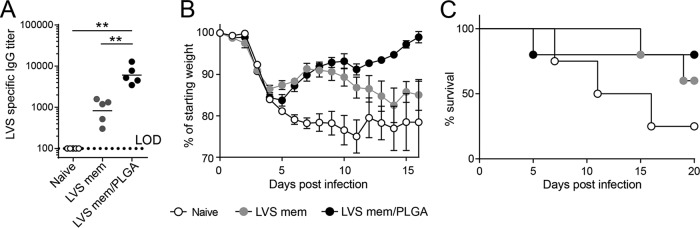
Francisella tularensis is the causative agent of tularemia and a potential bioterrorism agent. In the present study, we isolated, identified, and quantified the proteins present in the membranes of the virulent type A strain, Schu S4, and the attenuated type B strain, LVS (live vaccine strain). Spectral counting of mass spectrometric data showed enrichment for membrane proteins in both strains. Mice vaccinated with whole LVS membranes encapsulated in poly (lactic-co-glycolic acid) (PLGA) nanoparticles containing the adjuvant polyinosinic-polycytidylic acid [poly(I·C)] showed significant protection against a challenge with LVS compared to the results seen with naive mice or mice vaccinated with either membranes or poly(I·C) alone. The PLGA-encapsulated Schu S4 membranes with poly(I·C) alone did not significantly protect mice from a lethal intraperitoneal challenge with Schu S4; however, this vaccination strategy provided protection from LVS challenge. Mice that received the encapsulated Schu S4 membranes followed by a booster of LVS bacteria showed significant protection with respect to a lethal Schu S4 challenge compared to control mice. Western blot analyses of the sera from the Schu S4-vaccinated mice that received an LVS booster showed four immunoreactive bands. One of these bands from the corresponding one-dimensional (1D) SDS-PAGE experiment represented capsule. The remaining bands were excised, digested with trypsin, and analyzed using mass spectrometry. The most abundant proteins present in these immunoreactive samples were an outer membrane OmpA-like protein, FopA; the type IV pilus fiber building block protein; a hypothetical membrane protein; and lipoproteins LpnA and Lpp3. These proteins should serve as potential targets for future recombinant protein vaccination studies.IMPORTANCE The low infectious dose, the high potential mortality/morbidity rates, and the ability to be disseminated as an aerosol make Francisella tularensis a potential agent for bioterrorism. These characteristics led the Centers for Disease Control (CDC) to classify F. tularensis as a Tier 1 pathogen. Currently, there is no vaccine approved for general use in the United States.
Keywords: Franscisella tularensis; immunity; inner membrane proteins; outer membrane proteins; proteomics.
Copyright © 2017 Post et al.
Figures


Similar articles
-
Monophosphoryl Lipid A Enhances Efficacy of a Francisella tularensis LVS-Catanionic Nanoparticle Subunit Vaccine against F. tularensis Schu S4 Challenge by Augmenting both Humoral and Cellular Immunity.Clin Vaccine Immunol. 2017 Mar 6;24(3):e00574-16. doi: 10.1128/CVI.00574-16. Print 2017 Mar. Clin Vaccine Immunol. 2017. PMID: 28077440 Free PMC article.
-
Live Attenuated Tularemia Vaccines for Protection Against Respiratory Challenge With Virulent F. tularensis subsp. tularensis.Front Cell Infect Microbiol. 2018 May 15;8:154. doi: 10.3389/fcimb.2018.00154. eCollection 2018. Front Cell Infect Microbiol. 2018. PMID: 29868510 Free PMC article. Review.
-
Differential ability of novel attenuated targeted deletion mutants of Francisella tularensis subspecies tularensis strain SCHU S4 to protect mice against aerosol challenge with virulent bacteria: effects of host background and route of immunization.Vaccine. 2010 Feb 17;28(7):1824-31. doi: 10.1016/j.vaccine.2009.12.001. Epub 2009 Dec 16. Vaccine. 2010. PMID: 20018266 Free PMC article.
-
Novel catanionic surfactant vesicle vaccines protect against Francisella tularensis LVS and confer significant partial protection against F. tularensis Schu S4 strain.Clin Vaccine Immunol. 2014 Feb;21(2):212-26. doi: 10.1128/CVI.00738-13. Epub 2013 Dec 18. Clin Vaccine Immunol. 2014. PMID: 24351755 Free PMC article.
-
Francisella tularensis vaccines.FEMS Immunol Med Microbiol. 2007 Apr;49(3):315-23. doi: 10.1111/j.1574-695X.2007.00219.x. Epub 2007 Feb 22. FEMS Immunol Med Microbiol. 2007. PMID: 17316369 Review.
Cited by
-
Development, Phenotypic Characterization and Genomic Analysis of a Francisella tularensis Panel for Tularemia Vaccine Testing.Front Microbiol. 2021 Aug 11;12:725776. doi: 10.3389/fmicb.2021.725776. eCollection 2021. Front Microbiol. 2021. PMID: 34456897 Free PMC article.
-
A subunit vaccine against pneumonia: targeting Streptococcus pneumoniae and Klebsiella pneumoniae.Netw Model Anal Health Inform Bioinform. 2023;12(1):21. doi: 10.1007/s13721-023-00416-3. Epub 2023 Apr 19. Netw Model Anal Health Inform Bioinform. 2023. PMID: 37096010 Free PMC article.
-
Immunogenicity of trimeric autotransporter adhesins and their potential as vaccine targets.Med Microbiol Immunol. 2020 Jun;209(3):243-263. doi: 10.1007/s00430-019-00649-y. Epub 2019 Dec 1. Med Microbiol Immunol. 2020. PMID: 31788746 Free PMC article. Review.
-
A Metabolic Labeling Strategy for Relative Protein Quantification in Clostridioides difficile.Front Microbiol. 2018 Oct 16;9:2371. doi: 10.3389/fmicb.2018.02371. eCollection 2018. Front Microbiol. 2018. PMID: 30386308 Free PMC article.
-
Comparative evaluation of protective immunity against Francisella tularensis induced by subunit or adenovirus-vectored vaccines.Front Cell Infect Microbiol. 2023 May 25;13:1195314. doi: 10.3389/fcimb.2023.1195314. eCollection 2023. Front Cell Infect Microbiol. 2023. PMID: 37305410 Free PMC article.
References
-
- CDC 2012. Possession, use, and transfer of select agents and toxins; biennial review. Final rule. Fed Regist 77:61083–61115. - PubMed
MeSH terms
Substances
Grants and funding
LinkOut - more resources
Full Text Sources
Other Literature Sources
