. 2017 Oct 10;8(1):842.
doi: 10.1038/s41467-017-01011-7.

Stress-induced plasticity of dynamic collagen networks

Affiliations

Stress-induced plasticity of dynamic collagen networks

Jihan Kim et al. Nat Commun. .

Abstract

The structure and mechanics of tissues is constantly perturbed by endogenous forces originated from cells, and at the same time regulate many important cellular functions such as migration, differentiation, and growth. Here we show that 3D collagen gels, major components of connective tissues and extracellular matrix (ECM), are significantly and irreversibly remodeled by cellular traction forces, as well as by macroscopic strains. To understand this ECM plasticity, we develop a computational model that takes into account the sliding and merging of ECM fibers. We have confirmed the model predictions with experiment. Our results suggest the profound impacts of cellular traction forces on their host ECM during development and cancer progression, and suggest indirect mechanical channels of cell-cell communications in 3D fibrous matrices.The structure and mechanics of tissues is constantly perturbed by endogenous forces originated from cells. Here the authors show that 3D collagen gels, major components of connective tissues and extracellular matrix, are significantly and irreversibly remodelled by cellular traction forces and by macroscopic strains.


Conflict of interest statement

The authors declare no competing financial interests.

Figures

Fig. 1
Fig. 1
Cell traction forces irreversibly induce the formation of collagen bundles. a Reconstructed streamlines showing the spatial-temporal profile of the cell-induced matrix deformation. The deformation field from frame to frame is calculated via reflectance particle image velocimetry. Color code (blue to red) is linearly proportional to maturation time 0−30 min). b Confocal reflection image of the collagen matrix showing a collagen bundle (arrow) between two MDA-MB-231 cells. c Collagen bundles simultaneously form between multiple cell pairs. Red: GFP-labeled MDA-MB-231 cells. Green: reflectance image of collagen fibers. d The relative reflectance intensity ΔF/F of collagen bundles compared to the background. e ΔF/F of collagen bundles after disrupting the cell traction forces by Cytochalasin-D treatment. In d, e, ~30 cell pairs are sampled for each maturation time. The thick black lines, box edges and whiskers represent the median, first/third quartiles, and lower/upper 5% values, respectively. ANOVA and Fisher’s least significant difference procedure is used to evaluate the difference of ΔF/F corresponding to different maturation times. *p < 0.05, **p < 0.01. Differences between non-labeled pairs are not significant. f Fraction of permanent collagen bundles F perm as a function of maturation time. Scale bar: 20 μm
Fig. 2
Fig. 2
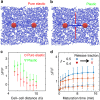
Simulation of collagen bundle formation by contracting cell pairs. a The network configuration in an elastic model (without any sliding or merging events). b The network configuration predicted by our plastic model. c The relative increase of fiber density ΔF/F of collagen bundles compared with the background at varying cell−cell distance. Here the cell−cell distance α is normalized by the cell size α. Red: plastic deformation with sliding events. Blue: pure elastic response of the network. At any given distance, the results from elastic (T = 0 min) and plastic (T = 15 min) are statistically distinct (t-test, p = 0.0007, N = 8). d The irreversibility of a collagen bundle depends on both cell contractility (β) and maturation time T d. Here the irreversibility is characterized by ΔF/F after the cell traction force is released. The cell−cell distance is fixed at d = 7a. Error bars in c, d: mean ± SD, obtained from eight different realizations
Fig. 3
Fig. 3
Bulk relaxation kinetics of collagen matrices. a The normalized elastic energy per fiber 〈H〉 over the course of relaxation of a model network. Black: sum of bending and stretching energy. Red: bending energy. Blue: stretching energy. All three curves are normalized by the total energy per fiber at t = −20 min. The network is sheared to 20% at t = −20 min, and released at t = 0. Inset: The network configuration after 20 min of relaxation (t = 1200 s). The fibers are color-coded according to the bending energy per unit length of each fiber H b, normalized by the ensemble average 〈H b〉. b Simulated strain decay kinetics with 20% initial strain and varying dwell times T d = 1, 2, 6, 10, 16, and 20 min. The dashed lines are fits to a single exponential. c Experiments show strain relaxation kinetics ε(t)−ε(∞) depend on the initial strain, and at small initial strains, the relaxation follows a single exponential function. Here ε(∞) is approximated by the strain measured after 15 min of relaxation, Supplementary Fig. 16 for results with extended relaxation time. d Experiments show strain relaxation kinetics depends on the dwell time T d. Colors of the symbols (blue to green) correspond to the increasing dwell time of 1, 2, 4, 7, 10, 15, and 20 min. Red lines are fit to double-exponential functions ε(t) = a exp(−t/τ v) + b exp(−t/τ p) + ε r. Here τ v is independent of dwell time T d, τ p, and ε r are allowed to vary with T d. Inset: zoom-in to the initial phase of the relaxation. e The plastic time scale τ p as a function of dwell time T m. f The residual strain ε r as a function of dwell time T d. Error bars in e, f are means and standard deviations from eight different samples
Fig. 4
Fig. 4
The micromechanics of collagen ECM in the vicinity of cell-induced collagen bundles after traction forces are released. a The confocal reflection image and directional compliance given by five probe particles around a collagen bundle in a typical experiment. The compliance is scaled linearly into real space such that an isotropic response of 0.5 Pa−1 would be plotted as a ring with the size of the bottom right circle. Magenta dots: experimentally measured directional compliance. Red circles: the compliance ellipse, i.e., the elliptical fit to the magenta dots. White dashed lines: outlines of MDA-MB-231 cells after Cytocytochalasin-D treatment. Green line: the location of collagen bundle. Scale bar: 50 μm. b The aspect ratios of the compliance ellipses at varying particle-to-bundle distances d. Symbols of different colors correspond to results measured around different bundles. We divide all the data into three groups d < 25 μm, 25 ≤ d < 50 μm, and 50 ≤ d < 75 μm. Error bars represent the mean and standard deviations of each group. ANOVA analysis shows that the aspect ratios close to the collagen bundles (d < 25 μm) are significantly higher than the values further away

Similar articles

Cited by

References

    1. Frantz C, Stewart KM, Weaver VM. The extracellula matrix at a glance. J. Cell Sci. 2010;123:4195. doi: 10.1242/jcs.023820. - DOI - PMC - PubMed
    1. Lu P, Weaver VM, Werb Z. The extracellular matrix: a dynamic niche in cancer progression. J. Cell Biol. 2012;196:395. doi: 10.1083/jcb.201102147. - DOI - PMC - PubMed
    1. Narayanan AS, Page RC, Swanson J. Collagen synthesis by human fibroblasts. regulation by transforming growth factor-β in the presence of other inflammatory mediators. Biochem. J. 1989;260:463. doi: 10.1042/bj2600463. - DOI - PMC - PubMed
    1. Mott JD, Werb Z. Regulation of matrix biology by matrix metalloproteinases. Curr. Opin. Cell Biol. 2004;16:558. doi: 10.1016/j.ceb.2004.07.010. - DOI - PMC - PubMed
    1. Egeblad M, Rasch MG, Weaver VM. Dynamic interplay between the collagen scaffold and tumor evolution. Curr. Opin. Cell Biol. 2010;22:697. doi: 10.1016/j.ceb.2010.08.015. - DOI - PMC - PubMed

Publication types

LinkOut - more resources