. 2017 Oct 10;7(1):12912.
doi: 10.1038/s41598-017-13141-5.

The human blood parasite Schistosoma mansoni expresses extracellular tegumental calpains that cleave the blood clotting protein fibronectin

Affiliations

The human blood parasite Schistosoma mansoni expresses extracellular tegumental calpains that cleave the blood clotting protein fibronectin

Qiang Wang et al. Sci Rep. .

Abstract

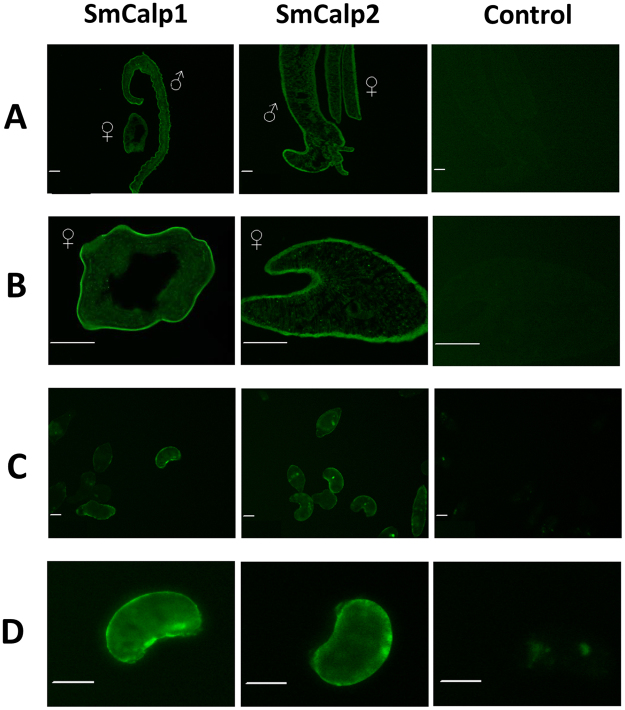
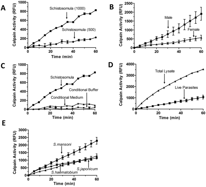
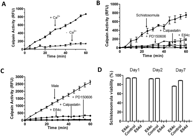
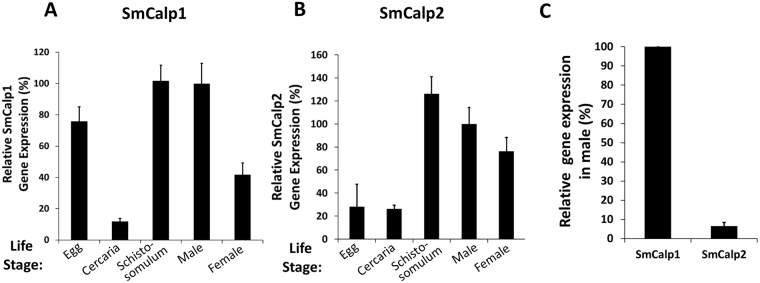
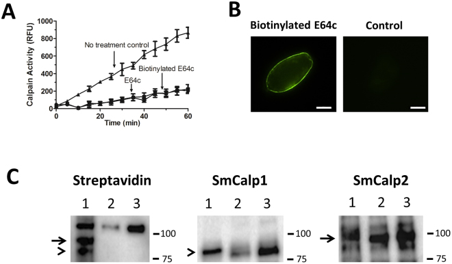
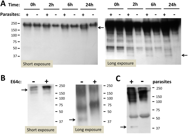
Schistosomes are intravascular, parasitic flatworms that cause debilitating disease afflicting >200 million people. Proteins expressed at the host-parasite interface likely play key roles in modifying the worm's local environment to ensure parasite survival. Proteomic analysis reveals that two proteases belonging to the calpain family (SmCalp1 and SmCalp2) are expressed in the Schistosoma mansoni tegument. We have cloned both; while highly conserved in domain organization they display just 31% amino acid sequence identity. Both display high relative expression in the parasite's intravascular life forms. Immunolocalization and activity based protein profiling experiments confirm the presence of the enzymes at the host-parasite interface. Living parasites exhibit surface calpain activity that is blocked in the absence of calcium and in the presence of calpain inhibitors (E64c, PD 150606 and calpastatin). While calpains are invariably reported to be exclusively intracellular (except in diseased or injured tissues), our data show that schistosomes display unique, constitutive, functional extracellular calpain activity. Furthermore we show that the worms are capable of cleaving the host blood clotting protein fibronectin and that this activity can be inhibited by E64c. We hypothesize that SmCalp1 and/or SmCalp2 perform this cleavage function to impede blood clot formation around the worms in vivo.


Conflict of interest statement

The authors declare that they have no competing interests.

Figures

Figure 1
Figure 1
SmCalp1 and SmCalp2 gene and protein organization. (A) Diagrammatic representation of the gene for SmCalp1 (top) and SmCalp2 (bottom). Exons are depicted in red and are numbered above each gene. Scale numbers below each gene represent kilobases (K). (B) Diagrammatic representation of S. mansoni chromosome 1 showing the location of the genes for SmCalp1 and SmCalp2. (C) Domain structure of SmCalp1, SmCalp2 and human calpain CAPN1. PC1 (grey) and PC2 (red) represent the protease core domain; CBSW domain is depicted in light blue and the penta-EF hand domain in green (containing five helix-loop-helix motifs (orange)). The letters and numbers on top of each PC1 and PC2 domain represent the positions of conserved active site amino acid residues. (D) Unrooted phylogenetic tree of selected calpains generated by neighbor joining with Accelrys Gene software. The scale bar represents the number of amino acid differences per unit length. Designations (and accession numbers) are as follows: S. mansoni Calp1 (SmCalp1; AAA29857.1); S. haematobium Calp 1 (ShCalp1, BAF62290.1); S. japonicum Calp1 (SjCalp1, BAA74718.1); Clonorchis sinensis Calp 1 (Cs1, GAA52477.1); Echinococcus granulosus Calp1 (Eg1, CDS23506.1); S. mansoni Calp2 (SmCalp2; MF590064); S. haematobium Calp2 (ShCalp2, XP_012791984.1); S. japonicum Calp2 (SjCalp2, MF590065); Clonorchis sinensis Calp2 (Cs2, GAA295577.2); Echinococcus granulosus Calp2 (Eg2, CDS20315.1); Drosophila melanogaster CalpB (Dm CalpB, NP_524016.4); Human calpain 9 (Hs CAPN9, NP_006606); Human calpain 2 (Hs CAPN2, NP_001739), Human calpain 1 (Hs CAPN1, NP_005177).
Figure 2
Figure 2
Immunolocalization of SmCalp1 and SmCalp2 in different S. mansoni life stages. (A) Longitudinal section of adult male and female parasites. (B) Cross section of adult female parasites. Scale bar in A and B represents 50 µm. (C,D) Whole 7-day cultured schistosomula; scale bar represents 25 µm. Preparations were probed with anti-SmCalp1 antibodies (left panel), anti-SmCalp2 antibodies (center panel) or no primary antibody (Control, right panel).
Figure 3
Figure 3
Measuring calpain activity in living schistosomes. (A) Calpain activity detected in live schistosomula (500 or 1000, as indicated) over time. (B) Calpain activity in living individual adult male (square,>10 individuals) and female (triangle,>10 individuals) parasites over time. (C) Calpain activity in 1000 live schistosomula (squares, groups of >5), compared to that seen in either conditional buffer (i.e. buffer that had contained 1,000 schistosomula for 1 h, up triangles) or in conditional medium (i.e. medium that had contained 1,000 schistosomula for 3 days, down triangles). (D) Calpain activity detected in individual living adult male parasites (squares) compared to that detected in total lysates of individual males (triangles). (E) Calpain activity in individual adult males of Schistosoma mansoni, S. japonicum, and S. haematobium (as indicated). All data are presented as relative fluorescence units (RFU, mean+/−SD, n ≥ 3). Calpain activity is given in relative fluorescence units (RFU). Fluorescence is generated following substrate cleavage and is measured at excitation/emission of 320/480.
Figure 4
Figure 4
Characterization of schistosome surface calpain activity. (A) Calpain activity measured in 1000 live schistosomula in the presence ( + ) or absence (−) of 3 mM Ca2+. Calpain activity detected in 1,000 living schistosomula (B) or individual male worms (C) in the presence or absence of the calpain inhibitors (E64c, calpastatin and PD150606, as indicated). Parasites were pre-incubated with inhibitor for 20 min before the addition of substrate. All activity data are presented as relative fluorescence units (RFU, mean+/−SD, n ≥ 3) where fluorescence is generated following substrate cleavage and is measured at excitation/emission 320/480. (D) Analysis of schistosomula viability after incubation in the presence of inhibitor (E64c or E64d, 100 µM) for 1, 2 or 7 days (as indicated). Data are shown relative to day 0, control parasite viability, set at 100%.
Figure 5
Figure 5
Expression of SmCalp1 and SmCalp2 in different S. mansoni life stages. Relative gene expression of SmCalp1 (A) and SmCalp2 (B) in the life stages indicated. All values are relative to males (set at 100%) (Mean+/− SD, n = 3). (C) Relative gene expression of SmCalp1 versus SmCalp2 in adult males (Mean+/− SD, n = 4). SmCalp1 gene expression is significantly higher than that of SmCalp2 (p < 0.0001, Students t test).
Figure 6
Figure 6
Activity based protein profiling of schistosome tegumental calpains. (A) Calpain activity in live adult male worms that were treated with E64c or biotinylated E64c or were untreated. Data are presented as relative fluorescence units (RFU) where fluorescence is derived following substrate cleavage and is measured at excitation/emission 320/480. Two-way ANOVA analysis were used to compare values; values from the no treatment control group differ significantly from either of the E64c treatment groups (p < 0.001, n = 5). (B) Localization of biotinylated E64c at the surface of a representative live schistosomulum (left panel); No staining is seen in the absence of biotinylated E64c treatment (Control, right panel). Scale bars represent 50 µm. (C) Western blot analysis of extracts of adult worms exposed to biotinylated E64c (lanes 1), E64c (lanes 2) or untreated control (lanes 3) worms. The blot was probed with streptavidin-HRP, (left panel) to detect biotinylated proteins, or with anti-SmCalp1 antibody (center) to detect SmCalp1, or with anti-SmCalp2 antibody (right panel) to detect SmCalp2. SmCalp blots were then probed with HRP-labeled secondary anti-antibody. The arrowhead indicates the position of migration of SmCalp1 and the arrow indicates the position of migration of SmCalp2. A non-specific streptavidin binding protein is detected at the top of all lanes in the “Streptavidin” panel (left). Numbers at right represent the positions of migration of molecular weight markers in kDa.
Figure 7
Figure 7
Fibronectin cleavage in the presence of schistosomes. (A) Biotinylated fibronectin was incubated in the presence (+) or absence (−) of schistosomula for different time periods (0, 2, 6 or 24 h, as indicated). At each time point aliquots were resolved by SDS-PAGE, blotted to PVDF membrane and probed with streptavidin-HRP. Short exposure of the membrane (left) shows appearance of a fibronectin degradation product (at ~180 kDa, arrow) only in the presence of parasites. Longer exposure (right) shows the concurrent appearance of a ~40 kDa fibronectin degradation product in the same preparations (arrow, right). Numbers at left represent the positions of migration of molecular weight markers, kDa. (B) Parasites were incubated with fibronectin in the presence (+) or absence (−) of the non-cell-permeable calpain inhibitor E64c. After 6 h, aliquots were recovered and resolved by SDS-PAGE, blotted to PVDF membrane and probed with streptavidin-HRP. It is clear that the presence of E64c impedes generation of both the~180 kDa high molecular weight (left, arrow) and the ~40 kDa lower molecular weight (right, arrow) fibronectin cleavage products. Numbers at right represent the positions of migration of molecular weight markers, kDa. (C) Schistosomula were incubated with mouse plasma for 6 h. An aliquot was recovered and resolved by SDS-PAGE, blotted to PVDF membrane and probed with anti-murine fibronectin antibody. A ~40 kDa band is detected (arrow, left lane) that is not seen in control plasma incubated in the absence of parasites (right lane). Numbers at right represent the positions of migration of molecular weight markers, kDa.

Similar articles

Cited by

References

    1. Gryseels B, Polman K, Clerinx J, Kestens L. Human schistosomiasis. Lancet. 2006;368:1106–1118. doi: 10.1016/S0140-6736(06)69440-3. - DOI - PubMed
    1. Steinmann P, Keiser J, Bos R, Tanner M, Utzinger J. Schistosomiasis and water resources development: systematic review, meta-analysis, and estimates of people at risk. Lancet Infect Dis. 2006;6:411–425. doi: 10.1016/S1473-3099(06)70521-7. - DOI - PubMed
    1. King CH, Dickman K, Tisch DJ. Reassessment of the cost of chronic helmintic infection: a meta-analysis of disability-related outcomes in endemic schistosomiasis. Lancet. 2005;365:1561–1569. doi: 10.1016/S0140-6736(05)66457-4. - DOI - PubMed
    1. King CH, Dangerfield-Cha M. The unacknowledged impact of chronic schistosomiasis. Chronic Illn. 2008;4:65–79. doi: 10.1177/1742395307084407. - DOI - PubMed
    1. Keating JH, Wilson RA, Skelly PJ. No overt cellular inflammation around intravascular schistosomes in vivo. J Parasitol. 2006;92:1365–1369. doi: 10.1645/GE-864R.1. - DOI - PubMed

Publication types