. 2017 Oct 10;7(1):12931.
doi: 10.1038/s41598-017-12788-4.

6E11, a highly selective inhibitor of Receptor-Interacting Protein Kinase 1, protects cells against cold hypoxia-reoxygenation injury

Affiliations

6E11, a highly selective inhibitor of Receptor-Interacting Protein Kinase 1, protects cells against cold hypoxia-reoxygenation injury

C Delehouzé et al. Sci Rep. .

Abstract

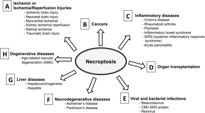
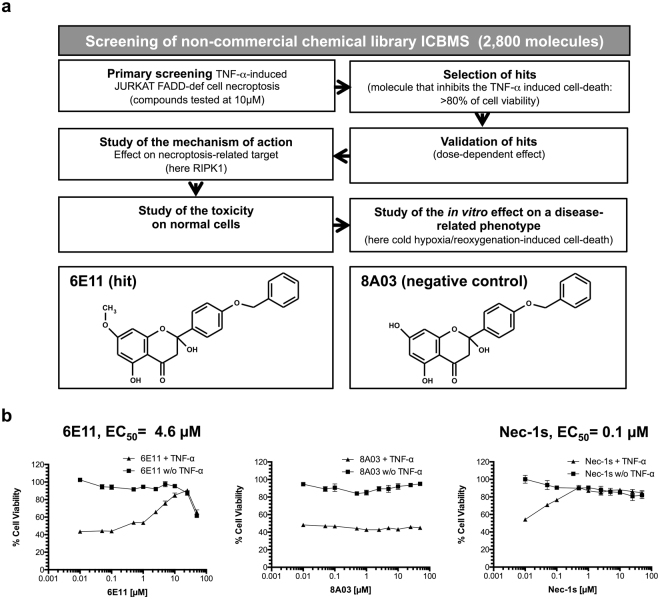
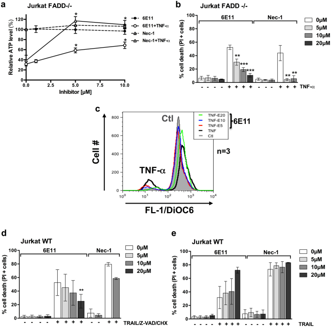
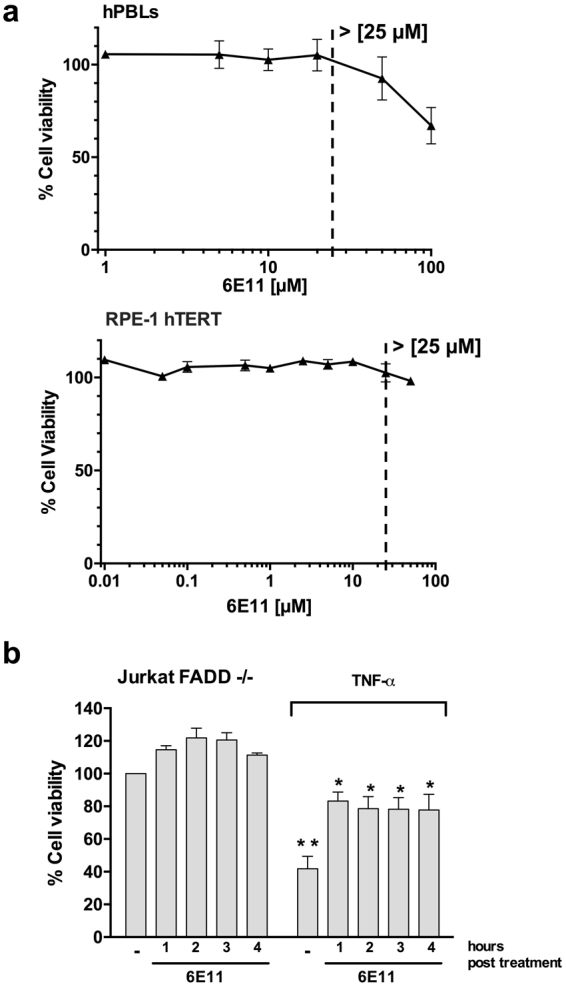
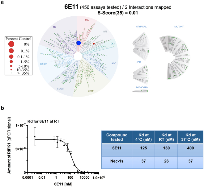
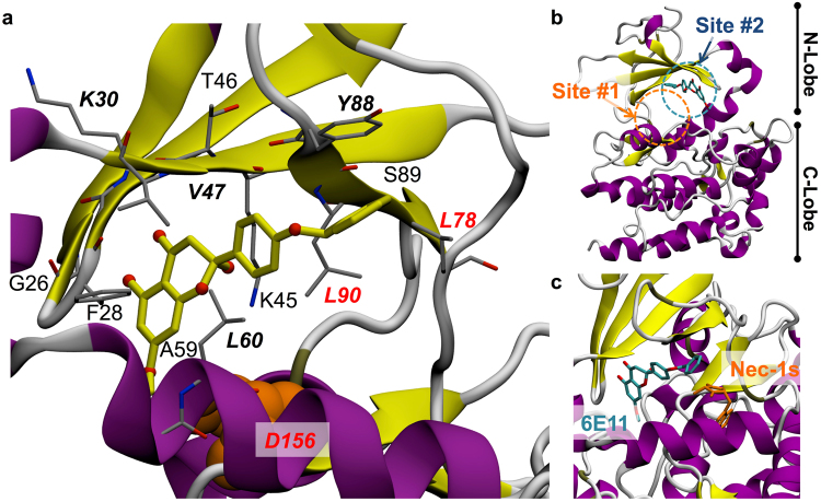
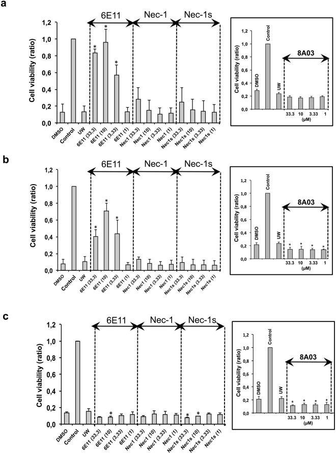
Necroptosis is a programmed cell death pathway that has been shown to be of central pathophysiological relevance in multiple disorders (hepatitis, brain and cardiac ischemia, pancreatitis, viral infection and inflammatory diseases). Necroptosis is driven by two serine threonine kinases, RIPK1 (Receptor Interacting Protein Kinase 1) and RIPK3, and a pseudo-kinase MLKL (Mixed Lineage Kinase domain-Like) associated in a multi-protein complex called necrosome. In order to find new inhibitors for use in human therapy, a chemical library containing highly diverse chemical structures was screened using a cell-based assay. The compound 6E11, a natural product derivative, was characterized as a positive hit. Interestingly, this flavanone compound: inhibits necroptosis induced by death receptors ligands TNF-α (Tumor Necrosis Factor) or TRAIL (TNF-Related Apoptosis-Inducing Ligand); is an extremely selective inhibitor, among kinases, of human RIPK1 enzymatic activity with a nM Kd; has a non-ATP competitive mode of action and a novel putative binding site; is weakly cytotoxic towards human primary blood leukocytes or retinal pigment epithelial cells at effective concentrations; protects human aortic endothelial cells (HAEC) from cold hypoxia/reoxygenation injury more effectively than necrostatin-1 (Nec-1) and Nec-1s. Altogether, these data demonstrate that 6E11 is a novel potent small molecular inhibitor of RIPK1-driven necroptosis.


Conflict of interest statement

The authors declare that they have no competing interests.

Figures

Figure 1
Figure 1
Impact of RIPK1-dependent necroptosis in human diseases. The comprehensive list of the references can be found as Supplementary Table S1.
Figure 2
Figure 2
Characterization of hit compound 6E11 as new necroptosis inhibitor. (a) Workflow of the cell-based screening of ICBMS chemical library for the selection of new inhibitors of necroptosis. Among 2,800 compounds, 6E11, was selected as the more potent inhibitor of TNF-α-induced necroptosis in human FADD-deficient Jurkat T cells. The chemical structures of 6E11 and its negative control (8A03) are depicted above the workflow. The primary screening is performed in monoplicate. The negative control was not detected during the screening campaign. (b) Dose-dependent protection of 6E11 against TNF-α-induced Jurkat FADD deficient cell necroptosis. After a 24-h incubation of the cells with or without (w/o) TNF-α and increasing concentrations of tested compounds, the effect on the cell viability was evaluated by MTS reduction assay. The cells were treated only with the tested compound to evaluate its putative toxicity. The values were normalized as a percentage of cell viability, considering 100% viable cells in the control treated with DMSO (n = 3, mean ± SD).
Figure 3
Figure 3
6E11 inhibits death receptor-induced necroptosis, but not apoptosis. (a) Human FADD-deficient Jurkat T cells were treated or not with TNF-α (10 ng/ml) in presence or not of increasing concentrations of 6E11 (0, 1, 5, 10 µM) or Nec-1 (0, 5, 10 µM) for 18 hours. Intracellular ATP levels were measured with the CellTiter-Glo® Luminescent Cell Viability Assay (n = 3, mean ± SEM, *P < 0.05) (EC50 ~6 µM). (b) Human FADD-deficient Jurkat T cells were treated or not with TNF-α (10 ng/ml) in presence or not of increasing concentrations of 6E11 (0, 5, 10, 20 µM) or Nec-1 (0, 5, 10 µM) for 18 hours. The percentage of cell death was determined by propidium iodide staining using flow cytometry (n = 3, mean ± SEM, **P < 0.01 and ***P < 0.001). (c) Human FADD-deficient Jurkat T cells were treated or not with TNF-α (10 ng/ml) in presence or not of increasing concentrations of 6E11 (0, 5, 10, 20 µM) for 18 hours. The mitochondrial transmembrane potential (MTP) was measured using the fluorescent dye DiOC6(3) and flow cytometry analysis. (d) 6E11 inhibits TRAIL-induced necroptosis. Wild-type Jurkat T cells were treated or not with TRAIL (10 ng/ml), Z-VAD (30 µM) and CHX (1 µg/ml) for 18 hours in presence or not of increasing concentrations of 6E11 (0, 5, 10, 20 µM) or 10 µM Nec-1. The percentage of cell death was determined by propidium iodide staining using flow cytometry (n = 3, mean ± SEM, **P < 0.01). (e) 6E11 does not inhibit TRAIL-induced apoptosis. Wild-type Jurkat T cells were treated or not with TRAIL (20 ng/ml) for 18 hours in presence or not of increasing concentrations of 6E11 (0, 5, 10, 20 µM) or Nec-1 (0, 5, 10, 20 µM). The percentage of cell death was determined by propidium iodide staining using flow cytometry (n = 3, mean ± SEM).
Figure 4
Figure 4
6E11 is not cytotoxic at inhibitory concentration and protects from necroptosis even after cell death initiation. (a) 24 hours after treatment with increasing concentrations of 6E11, cell viability was measured with a MTS assay to determine the toxicity of the compound towards either human PBL (upper panel) (n = 6 individuals, mean ± SEM) or human retina pigmented epithelial cells (RPE-1 hTERT) (lower panel) (n = 3, mean ± SD). (b) Viability was measured with a MTS assay in FADD-def Jurkat cells treated with TNF-α (10 ng/ml) followed by 10 µM 6E11 addition 1 to 4 h post necroptosis initiation (n = 4, mean ± SEM, *P < 0.05; **P < 0.01).
Figure 5
Figure 5
6E11 is a highly selective inhibitor of human kinase RIPK1. (a) Selectivity of 6E11 was assessed biochemically against a panel of 456 purified kinases (KINOMEscanSM Assay) at 10 µM and the compound was found to be highly selective for RIPK1. Only 0.15% of the initial amount of RIPK1 is still on the affinity matrix after competition with 6E11 (the full list of results is reported in Supplementary Table S2). S-Score35 = (number of non-mutant kinases with %Ctrl < 35)/(number of non-mutant kinases tested). (b) Measure of the binding constant for RIPK1/6E11 and RIPK1/Nec-1s interactions. Two 11-point 3-fold serial dilutions of 6E11 and Nec-1s were prepared in 100% DMSO in order to determine the binding constant (Kd). Kds are calculated at three various temperatures by measuring the amount of kinase captured on the solid support as a function of the test compound concentration (n = 2, mean ± range).
Figure 6
Figure 6
6E11 inhibits the enzymatic activity of RIPK1 with a non-ATP competitive mode of action. (a) RIPK1 was treated with 5, 10 or 50 µM of 6E11 to analyze the effect on the kinase autophosphorylation. Radioactive autophosphorylation assays were processed with [γ-32P] ATP at 30 µM final concentration. Necrostatin-1 (Nec-1) was used as an internal control. Coomassie blue staining was performed in order to estimate the total amount of protein loaded on polyacrylamide gel. Autophosphorylated RIPK1 band was visualized on radiographic film. % of RIPK1 activity inhibition are calculated through a ratio with DMSO, as a 0 percent reference. See Supplementary Fig. S2 for results of the full-length gel obtained at various exposure times. (b) ATP competition assay shows that inhibition of RIPK1 activity by 6E11 is not affected by ATP concentration. On the left panel, 0.25 or 1 mM of cold ATP were used to study the competition with 6E11. These assays are similar to those described in (a). The phosphorylation signal is modified by increasing doses of ATP due to the dilution of [γ-32P] ATP by “cold” non radiolabeled ATP. % of RIPK1 activity inhibition are calculated for each ATP concentration through a ratio with DMSO (-), as a 0 percent reference. See Supplementary Fig. S2 for results of the full-length gel. The RIPK1 catalytic activity was also monitored using myelin basic protein, MBP, as a substrate (right panel). GST-RIPK1 full length produced by baculovirus in Sf9 insect cells was assayed in the presence of increasing concentrations of 6E11. The kinase activities are expressed in % of maximal activity, i.e. measured in the absence of inhibitor. The IC50 values obtained at various ATP concentrations are reported in the table (n = 3, mean ± SD). (c) 6E11 is inactive on RIPK3 activity. GST-RIPK3 full length produced by baculovirus in Sf9 insect cells was assayed in the presence of 10 µM of inhibitor with [γ-33P] ATP and myelin basic protein, MBP, as a substrate. GSK’872 was used as RIPK3 reference inhibitor. Kinase activities are expressed in % of maximal activity, i.e. measured in the presence of DMSO (as a 100% reference).
Figure 7
Figure 7
Molecular model of the RIPK1-6E11 complex. (a) Close-up view of the putative interaction between 6E11 and surrounding residues in RIPK1 extracted from the best pose of docking simulation improved by 60 ns molecular dynamic trajectory (key-residues are shown in italic and bold, residues common to Nec-1s and 6E11 binding sites are in red). Asp156, one of the RIPK1 catalytic triad residues, but more slightly contacting 6E11, is shown in orange spheres to complete the description of the close environment of the ligand. (b) Overall structure of RIPK1 showing the putative binding site of 6E11 highlighted in blue dashed circle (site #2). The binding site (site #1) of Nec-1s (determined from the X-ray structure 4ITH) is marked with a orange dashed circle on the same panel. (c) Relative positions and orientations of each 6E11 and Nec-1s compounds shown in a close-up view.
Figure 8
Figure 8
6E11 protects HAEC from cold hypoxia reoxygenation injury when treatment occurs during cold hypoxia step (a), during both cold hypoxia and reoxygenation steps (b), but not when treatment occurs during the reoxygenation step (c). Increasing concentrations (1, 3.33, 10, 33.3 µM) of 6E11, Nec-1, Nec-1s or 8A03 were added to the UW preservation solution during hypoxia or/and in PBS – 2% FBS during reoxygenation. UW corresponds to cells treated with UW preservation solution. Controls are cells not subjected to this protocol and continuously oxygenated. DMSO are cells treated with drug vehicle. The ratio of cell viability evaluated by XTT test, is calculated in comparison to the untreated control (100% of cell viability). Data shown are mean ± SD, n = 3; ANOVA and Tukey’s Multiple Comparison Test; *P < 0.05 vs UW. See Supplementary Fig. S3 for a schematic representation of the protocol.

References

    1. Conrad M, Angeli JPF, Vandenabeele P, Stockwell BR. Regulated necrosis: disease relevance and therapeutic opportunities. Nat. Rev. Drug Discov. 2016;15:348–366. doi: 10.1038/nrd.2015.6. - DOI - PMC - PubMed
    1. Degterev A, et al. Chemical inhibitor of nonapoptotic cell death with therapeutic potential for ischemic brain injury. Nat. Chem. Biol. 2005;1:112–119. doi: 10.1038/nchembio711. - DOI - PubMed
    1. Degterev A, et al. Identification of RIP1 kinase as a specific cellular target of necrostatins. Nat. Chem. Biol. 2008;4:313–321. doi: 10.1038/nchembio.83. - DOI - PMC - PubMed
    1. Vanden Berghe T, Linkermann A, Jouan-Lanhouet S, Walczak H, Vandenabeele P. Regulated necrosis: the expanding network of non-apoptotic cell death pathways. Nat. Rev. Mol. Cell Biol. 2014;15-:135–147. doi: 10.1038/nrm3737. - DOI - PubMed
    1. Miao B, Degterev A. Methods to analyze cellular necroptosis. Methods Mol. Biol. 2009;559:79–93. doi: 10.1007/978-1-60327-017-5_6. - DOI - PubMed

Publication types

MeSH terms

Substances

LinkOut - more resources