Review
. 2017 Sep 22:8:737.
doi: 10.3389/fphys.2017.00737. eCollection 2017.

Adenosine: Direct and Indirect Actions on Gastric Acid Secretion

Affiliations
Review

Adenosine: Direct and Indirect Actions on Gastric Acid Secretion

Rosa M Arin et al. Front Physiol. .

Abstract

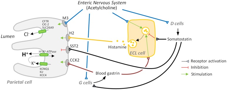
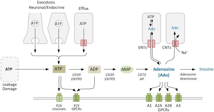
Composed by a molecule of adenine and a molecule of ribose, adenosine is a paradigm of recyclable nucleoside with a multiplicity of functions that occupies a privileged position in the metabolic and regulatory contexts. Adenosine is formed continuously in intracellular and extracellular locations of all tissues. Extracellular adenosine is a signaling molecule, able to modulate a vast range of physiologic responses in many cells and organs, including digestive organs. The adenosine A1, A2A, A2B, and A3 receptors are P1 purinergic receptors, G protein-coupled proteins implicated in tissue protection. This review is focused on gastric acid secretion, a process centered on the parietal cell of the stomach, which contains large amounts of H+/K+-ATPase, the proton pump responsible for proton extrusion during acid secretion. Gastric acid secretion is regulated by an extensive collection of neural stimuli and endocrine and paracrine agents, which act either directly at membrane receptors of the parietal cell or indirectly through other regulatory cells of the gastric mucosa, as well as mechanic and chemic stimuli. In this review, after briefly introducing these points, we condense the current body of knowledge about the modulating action of adenosine on the pathophysiology of gastric acid secretion and update its significance based on recent findings in gastric mucosa and parietal cells in humans and animal models.

Keywords: enteric nervous system; extracellular adenosine; gastric acid secretion regulation; gastric mucosa; purinergic signaling.


Figures

Figure 1
Figure 1
Major players in gastric acid secretion. Apical secretion of hydrochloric acid by the parietal cell requires three ion transport processes: an H+/K+-ATPase-mediated exchange of intracellular hydrogen ion for an extracellular potassium ion, and chloride secretion and potassium recycling, which are necessary to maintain electroneutrality and avoid luminal potassium depletion. Secretion is mainly regulated by activation: (i) of receptors for the secretagogues acetylcholine, histamine, and gastrin that are mainly secreted by the enteric nervous system, enterochromaffin-like (ECL) cells, and G cells, respectively, and (ii) of receptors for inhibitory somatostatin released by D cells.
Figure 2
Figure 2
Adenosine metabolism in the extracellular space. Extracellular adenosine mainly derives from the phosphohydrolysis of precursor nucleotides ATP, ADP, and AMP by ectonucleotidases CD39 and CD73. Adenosine levels also depend on the activity of equilibrative and concentrative nucleoside transporters ENTs and CNTs, that allow the nucleoside to cross the plasma membrane, and of adenosine deaminase that degrades irreversibly adenosine to inosine. Purinergic receptors comprise the ligand-gated ion channel P2X and the G protein-coupled receptors (GPCRs) P2Y for nucleotides, and the four GPCRs for adenosine (P1). ENTPD, family of ectonucleoside triphosphate diphosphohydrolases. Purinergic receptors (P1 and/or P2) and other proteins co-expressed on the membrane may form cell type-specific combinatorial signaling units.
Figure 3
Figure 3
The innervation of the different layers of the stomach wall is a potential adenosine source. Parasympathetic fibers innervate the myenteric and submucosal plexuses of the enteric nervous system and acetylcholine (ACh) is the major neurotransmitter, while sympathetic fibers innervate the same regions and the gastric mucosa, being norepinephrine (NE) the major neurotransmitter. Brown lines represent innervation between different regions of the intrinsic nervous system. ATP, which is co-released in all the synaptic contacts, can be enzymatically transformed into adenosine.

References

    1. Abbracchio M. P., Burnstock G. (1994). Purinoceptors: are there families of P2X and P2Y purinoceptors? Pharmacol. Ther. 64, 445–475. 10.1016/0163-7258(94)00048-4 - DOI - PubMed
    1. Adriaenssens A., Lam B. Y., Billing L., Skeffington K., Sewing S., Reimann F., et al. . (2015). A transcriptome-led exploration of molecular mechanisms regulating somatostatin-producing D-cells in the gastric Epithelium. Endocrinology 156, 3924–3936. 10.1210/en.2015-1301 - DOI - PMC - PubMed
    1. Ainz L. F., Gil-Rodrigo C. E., Gomez R., Malillos M., Requejo D., Gandarias J. M. (1989). Effects of various physiologic adenine derivatives on the secretion of acid in isolated gastric glands in rabbits. Rev. Esp. Fisiol. 45, 281–286. - PubMed
    1. Ainz L. F., Salgado C., Gandarias J. M., Gomez R., Vallejo A., Gil-Rodrigo C. E. (1993). P1(A2/Ra)-purinoceptors may mediate the stimulatory effect of adenosine and adenosine analogs on acid formation in isolated rabbit parietal cells. Pharmacol. Res. 27, 319–334. 10.1006/phrs.1993.1032 - DOI - PubMed
    1. Alumets J., Ekelund M., El Munshid H. A., Hakanson R., Loren I., Sundler F. (1979). Topography of somatostatin cells in the stomach of the rat: possible functional significance. Cell Tissue Res. 202, 177–188. 10.1007/BF00232233 - DOI - PubMed

LinkOut - more resources