. 2017 Sep 22:8:1192.
doi: 10.3389/fimmu.2017.01192. eCollection 2017.

IFN-γ-STAT1-iNOS Induces Myeloid Progenitors to Acquire Immunosuppressive Activity

Affiliations

IFN-γ-STAT1-iNOS Induces Myeloid Progenitors to Acquire Immunosuppressive Activity

Shu-Han Yang et al. Front Immunol. .

Abstract

Autoimmune diseases often induce dysregulated hematopoiesis with altered number and function of hematopoietic stem and progenitor cells (HSPCs). However, there are limited studies on the direct regulation of HSPCs on T cells, which are often detrimental to autoimmunity. Here, we found that in a murine model of Concanavalin A-induced autoimmune hepatitis, LSK (Lineage-Sca-1+c-Kit+)-like cells accumulated in liver, spleen, and bone marrow (BM), which were myeloid progenitors (Lineage-Sca-1-c-Kit+) that upregulated Sca-1 expression upon T cell-derived IFN-γ stimulation. Strikingly, BM LSK-like cells from mice induced by Con A to develop autoimmune hepatitis or alternatively myeloid progenitors from wild-type mice possessed strong in vitro suppressive ability. Their suppressive function depended on T cell-derived IFN-γ in a paracrine fashion, which induced STAT1 phosphorylation, inducible nitric oxide synthase expression, and nitric oxide production. Blocking IFN-γ/IFN-γ receptor interaction, knockout of STAT1, or iNOS inhibition abrogated their suppressive function. In addition, the suppressive function was independent of differentiation; mitomycin C-treated myeloid progenitors maintained T cell suppressive ability in vitro. Our data demonstrate a mechanism of inflammation induced suppressive function of myeloid progenitors, which may participate directly in suppressing T cell-mediated immunopathology.

Keywords: IFN-γ; STAT1; T cells; autoimmune disease; bone marrow; immunosuppression; inducible nitride oxide synthase; myeloid progenitors.


Figures
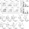
Figure 1
Figure 1
LSK-like cell accumulation during Con A-induced liver AIH. (A) Representative flow cytometry result of LSK cells in BM, liver, and spleen of 10-week-old wild type male mice 24 h after injection i.v. with PBS (n = 4) and 10 mg/kg Con A (n = 4). Numbers represent percentage of lineage cells. (B,C) Statistical analysis of (B) percentage and (C) number of LSK cells in BM, liver, and spleen of Con A- or PBS-treated mice. (D,E) LSK percentage of (D) BM, liver, spleen leukocytes, (E) serum IFN-γ, and alanine aminotransferase (ALT) level at 0, 6, 12, 24, 48, and 72 h after Con A treatment. (F) MHC-II molecule, PD-L1, and CD86 expression of BM LSK-like cells at different time points after Con A injection. (G) Percentage of Ki67+ BM LSK-like cells at different time point after Con A treatment. N = 3 per time point. *p < 0.05, **p < 0.01, ***p < 0.001. Data are shown in mean ± SEM.
Figure 2
Figure 2
WT LSK cells and LSK-like cells from Con A-treated mice inhibit T cell proliferation in vitro. (A,B) Proliferation of carboxyfluorescein succinimidyl ester-labeled OT-II T cells (5 × 104/well) cocultured with WT LSK or Con A BM LSK-like cells (1 × 104/well), respectively, for 72 h, in the presence of soluble OVA peptide 323–339 (1 µg/ml) and WT B cells (CD19+ cells from spleen of WT mice, 1 × 104/well). (C) Proliferation of OVA peptide and B cell-activated OT-II T cells cocultured with WT LSK cells. (D) Proliferation of OVA peptide and B cell-activated OT-II T cells cocultured with WT LSK or Con A BM LSK-like cells at indicated ratios. (E) The suppression ability of common myeloid progenitors (CMPs), granulocyte-monocyte progenitor (GMP), and megakaryocyte–erythrocyte progenitor subsets, respectively, sorted from WT BM LSK cells. (F) The suppression ability of liver and spleen LSK-like cells from mice treated with con A for 24 h. (G) Subset of liver, spleen, and BM LSK-like cells from Con A-treated mice at indicated time points. N = 3 for coculture experiments and data represent one of at least two independent experiments. *p < 0.05, **p < 0.01, ***p < 0.001. Data are shown in mean ± SEM.
Figure 3
Figure 3
LSK cells suppress T cell proliferation through iNOS activation. (A) Proliferation of OVA peptide and B cell-activated OT-II T cells cocultured with WT or H2Ab1−/− BM LSK cells. (B) Suppression ability of WT LSK cells in the presence of anti-PD-L1 antibody (5 µg/ml) and isotype control. (C,D) Suppression ability of (C) WT LSK cells and (D) Con A BM LSK-like cells in the presence of L-NMMA [total nitric oxide synthase (NOS) inhibitor], 1,400 W (iNOS inhibitor), or LNAME (eNOS inhibitor). Suppression experiments set as in this figure. (E) Expression of iNOS mRNA of WT LSK cocultured with OVA stimulated OT-II T cells after 0, 6, 12, 24, and 48 h. (F) Proliferation of OVA peptide and B cell (5 × 104)-activated OT-II T cells (2.5 × 105) cocultured with or without WT LSK cells (5 × 104) in the top or bottom chamber of transwell system. *p < 0.05, **p < 0.01, ***p < 0.001. N = 3 for coculture experiments and data represent one of at least two independent experiments. Data are shown in mean ± SEM.
Figure 4
Figure 4
IFN-γ boosts LSK cells suppressive potential via STAT-1–iNOS pathway. (A) Proliferation of OVA peptide and B cell-activated OT-II T cells cocultured with WT or GRKO LSK cells. (B) Suppression ability of WT LSK cells on the proliferation of OVA peptide and B cell-activated GKO OT-II T cells. Suppression experiments set as Figure 3. (C) Expression of iNOS mRNA of WT LSK cocultured with OVA-stimulated OT-II T cells or GKO OT-II T cells after 24 h. (D) Suppression ability of WT LSK cells on the proliferation of OVA and B cell-activated GKO OT-II T cells under extra IFN-γ administration at indicated concentration. (E) Suppression ability of WT LSK cells on the proliferation of OVA peptide and B cell-activated GKO OT-II T cells in the presence of OT-II T cells (1 × 104) or GKO OT-II T cells (1 × 104) as control. Suppression experiments set as Figure 3. (F) STAT1 phosphorylation of WT BM LSK cells and BM LSK-like cells from Con A-treated mice. (G) Proliferation of OVA peptide and B cell-activated OT-II T cells cocultured with WT or STAT1−/− LSK cells. (H) Expression of iNOS mRNA of WT LSK or STAT1−/− LSK cells cocultured with OVA-stimulated OT-II CD4+T cells after 24 h. N = 3 for coculture experiments and data represent one of at least two independent experiments. *p < 0.05, **p < 0.01, ***p < 0.001. Data are shown in mean ± SEM.
Figure 5
Figure 5
T cell suppressive ability of LSK cells is independent of differentiation. (A) Proliferation of WT LSK cells (5 × 104) cocultured with OT-II T cells or GKO OT-II T cells (5 × 104) under OVA (1 µg/ml) stimulation. (B) Differentiation of WT LSK cells (5 × 104) cocultured with OT-II T cells (5 × 104) under OVA (1 µg/ml) stimulation or not after 24, 48, and 72 h. (C) Proliferation of OVA and B cell-activated OT-II T cells cocultured with mitomycin C-treated WT LSK cells. (D) Analyses of the suppression ability of mitomycin C-treated WT LSK cells on OVA and B cell-activated OT-II T cells in the presence of 1,400 W. (E,F) Proliferation of OVA and B cell-activated OT-II T cells cocultured with (E) CD11b+ly6Ghi cells and (F) CD11b+ly6Chi cells from spleen of WT or Con A-treated mice. Suppression experiments set as Figure 3. *p < 0.05, **p < 0.01, ***p < 0.001. N = 3 for coculture experiments and data represent one of at least two independent experiments. Data are shown in mean ± SEM.

Similar articles

Cited by

References

    1. Morita Y, Ema H, Nakauchi H. Heterogeneity and hierarchy within the most primitive hematopoietic stem cell compartment. J Exp Med (2010) 207(6):1173–82.10.1084/jem.20091318 - DOI - PMC - PubMed
    1. Calvi LM, Link DC. The hematopoietic stem cell niche in homeostasis and disease. Blood (2015) 126(22):2443–51.10.1182/blood-2015-07-533588 - DOI - PMC - PubMed
    1. Massberg S, Schaerli P, Knezevic-Maramica I, Kollnberger M, Tubo N, Moseman EA, et al. Immunosurveillance by hematopoietic progenitor cells trafficking through blood, lymph, and peripheral tissues. Cell (2007) 131(5):994–1008.10.1016/j.cell.2007.09.047 - DOI - PMC - PubMed
    1. King KY, Goodell MA. Inflammatory modulation of HSCs: viewing the HSC as a foundation for the immune response. Nat Rev Immunol (2011) 11(10):685–92.10.1038/nri3062 - DOI - PMC - PubMed
    1. Laura M, Calvi DCL. The hematopoietic stem cell niche in homeostasis and disease. Blood (2015) 126(22):10.10.1182/blood-2015-07-533588 - DOI - PMC - PubMed