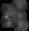
Retinal detachment with a break at pars plicata associated with congenital malformation of the lens-zonule-ciliary body complex
- PMID: 29018688
- PMCID: PMC5602713
- DOI: 10.1016/j.tjo.2014.06.003
Retinal detachment with a break at pars plicata associated with congenital malformation of the lens-zonule-ciliary body complex
Abstract
Retinal detachment with a break at the pars plicata associated with congenital malformation of lens-zonule-ciliary body complex is rare; most reports are of young Japanese male patients with atopic dermatitis. The present case report is the first to describe the condition in a Chinese patient with no atopic dermatitis or trauma history. A 22-year-old male presented with blurred vision in the left eye for 4 months. Fundus examination revealed shallow lower temporal retinal detachment. Further examination with scleral indentation under maximal pupil dilatation identified a break at the far periphery beyond the ora serrata and pars plana. Gonioscopy revealed a pars plicata break at the nonpigmented ciliary epithelium associated with congenital ciliary process hypoplasia and subtle lens defect at the same meridian. The retina was successfully reattached after segmental scleral buckling, cryopexy, and laser photocoagulation.
Keywords: malformation of the lens—zonule—ciliary body complex; pars plicata break; retinal detachment; scleral buckle.
Conflict of interest statement
Conflicts of interest: The authors have no potential financial or nonfinancial conflicts of interest to declare.
Figures


Similar articles
-
Retinal detachment with breaks in the pars plicata of the ciliary body.Am J Ophthalmol. 1989 Oct 15;108(4):349-55. doi: 10.1016/s0002-9394(14)73299-4. Am J Ophthalmol. 1989. PMID: 2801853
-
Bilateral retinal detachment with large breaks of pars plicata associated with coloboma lentis and ocular hypertension.Jpn J Ophthalmol. 1992;36(1):97-102. Jpn J Ophthalmol. 1992. PMID: 1635302
-
Retinal detachment in patients with atopic dermatitis. 5-year retrospective survey.Ophthalmologica. 1995;209(3):160-4. doi: 10.1159/000310604. Ophthalmologica. 1995. PMID: 7630624
-
[Variation of inflammatory reaction of ciliary body--harmony between clinic and basic science].Nippon Ganka Gakkai Zasshi. 2004 Dec;108(12):717-48; discussion 749. Nippon Ganka Gakkai Zasshi. 2004. PMID: 15656085 Review. Japanese.
-
Primary retinal detachment: scleral buckle or pars plana vitrectomy?Curr Opin Ophthalmol. 2006 Jun;17(3):245-50. doi: 10.1097/01.icu.0000193097.28798.fc. Curr Opin Ophthalmol. 2006. PMID: 16794436 Review.
Cited by
-
Ultrasound biomicroscopy-based diagnosis and cryotherapy for idiopathic rhegmatogenous retinal detachment secondary to pars plicata non-pigmented epithelium break: a case description.Quant Imaging Med Surg. 2025 Aug 1;15(8):7618-7623. doi: 10.21037/qims-2025-329. Epub 2025 Jul 25. Quant Imaging Med Surg. 2025. PMID: 40785879 Free PMC article. No abstract available.
References
-
- Long JC, Danielson RW. Traumatic detachment of retina and of pars ciliaris retinae. Am J Ophthalmol. 1953;36:515–516. - PubMed
-
- Hida T, Tano Y, Okinami S, Ogino N, Inoue M. Multicenter retrospective study of retinal detachment associated with atopic dermatitis. Jpn J Ophthalmol. 2000;44:407–418. - PubMed
-
- Iijima Y, Wagai K, Matsuura Y, Ueda M, Miyazaki I. Retinal detachment with breaks in the pars plicata of the ciliary body. Am J Ophthalmol. 1989;108:349–355. - PubMed
-
- Kusaka S, Ohashi Y. Retinal detachments with crescent-shaped retinal breaks in patients with atopic dermatitis. Retina. 1996;16:312–316. - PubMed
-
- Uemura A, Uto M. Bilateral retinal detachment with large breaks of pars plicata associated with coloboma lentis and ocular hypertension. Jpn J Ophthalmol. 1992;36:97–102. - PubMed
Publication types
LinkOut - more resources
Full Text Sources
Other Literature Sources
Miscellaneous

