Retinal complications associated with congenital optic disc anomalies determined by swept source optical coherence tomography
- PMID: 29018703
- PMCID: PMC5602127
- DOI: 10.1016/j.tjo.2015.05.003
Retinal complications associated with congenital optic disc anomalies determined by swept source optical coherence tomography
Abstract
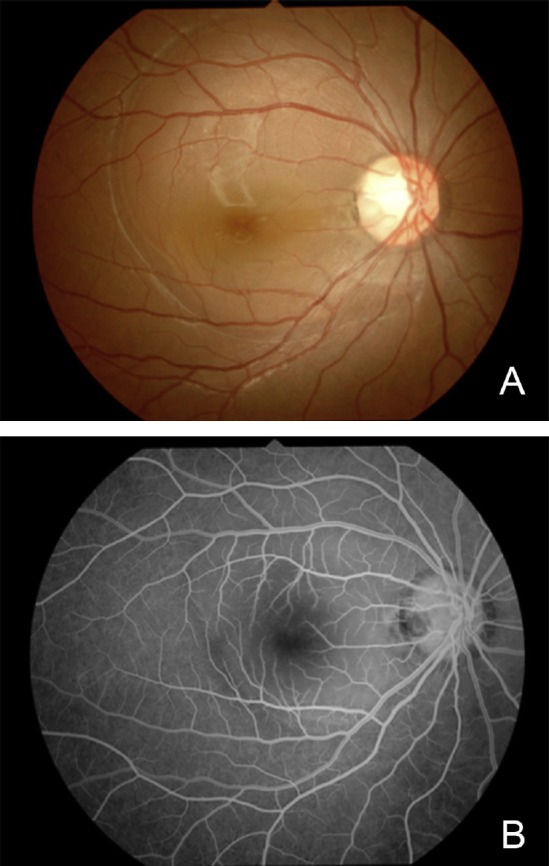
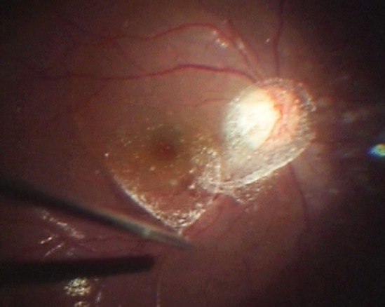
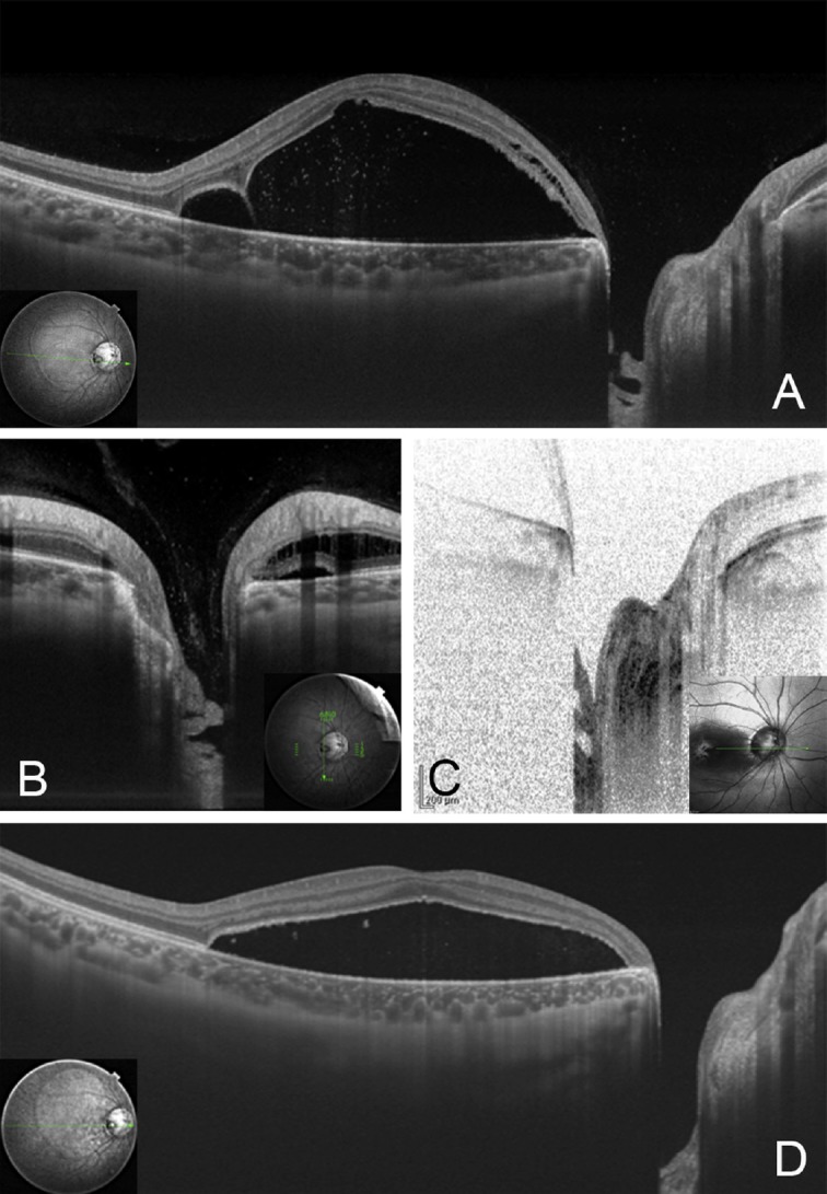
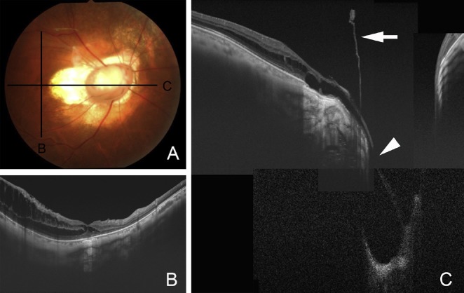
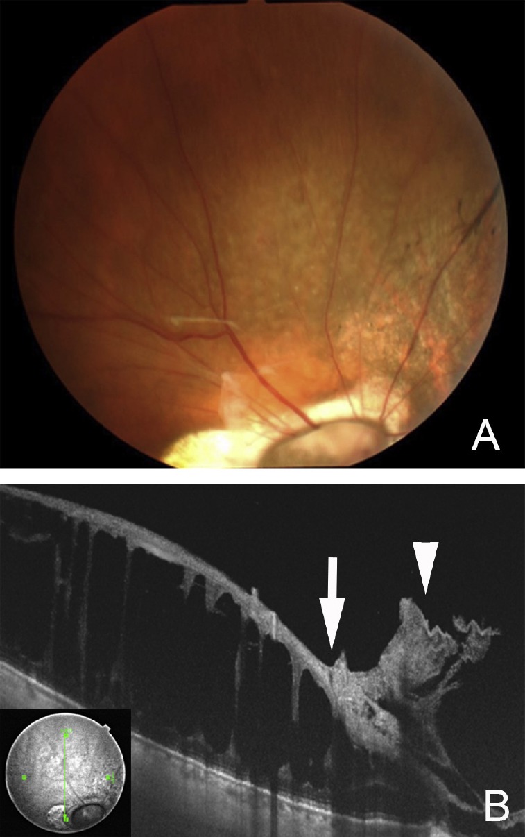
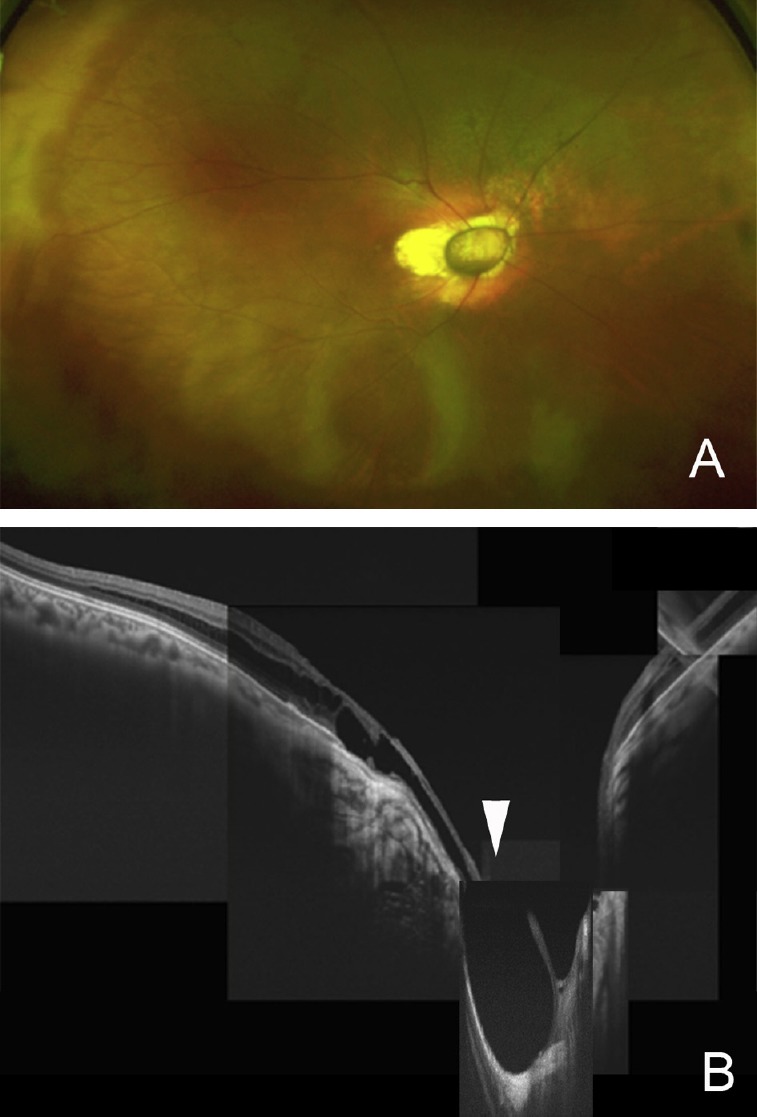
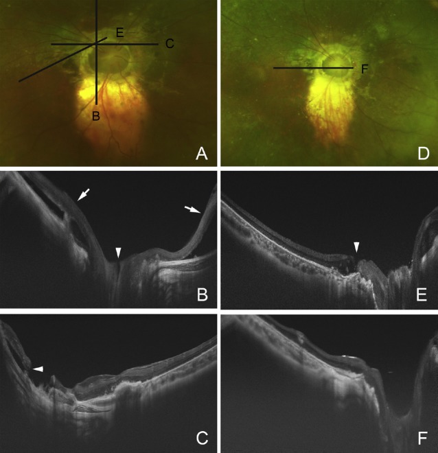
Optical coherence tomography has evolved over the past 2 decades to be an important ancillary method to evaluate diseases of the anterior and posterior segments of the eye. The more recent development of swept-source optical coherence tomography (SS-OCT) with a wavelength-tunable laser centered at 1050 nm and deeper imaging depth of 2.6 mm has enabled clinicians to evaluate congenital optic disc anomalies including optic disc pits, optic disc colobomas, and morning glory syndrome in more detail. The SS-OCT findings of the posterior precortical vitreous pocket, Cloquet's canal, lamina cribrosa that is torn from the peripapillary sclera, and the retrobulbar subarachnoid space immediately posterior to the highly reflective tissue lining the bottom of the excavation are presented. In addition, abnormal communications between the vitreous cavity and the subretinal and subarachnoid spaces in eyes with congenital optic disc anomalies are also reviewed. The retinal complications associated with congenital optic disc anomalies are treated by vitreous surgery, silicone oil tamponade, and peripapillary laser photocoagulation or scleral buckling. However, the surgical outcomes are limited and not entirely satisfactory. Analyses by SS-OCT of congenital optic disc anomalies should make the treatment correspond better with the pathological findings.
Keywords: coloboma; congenital optic disc anomaly; morning glory syndrome; optic disc pit; optical coherence tomography.
Conflict of interest statement
Conflict of interest: The author has no conflict of interest concerning in this study
Figures
References
-
- Bartz-Schmidt KU, Heimann K. Pathogenesis of retinal detachment associated with morning glory disc. Int Ophthalmol. 1995;19:35–38. - PubMed
-
- Coll GE, Chang S, Flynn TE, Brown GC. Communication between the subretinal space and the vitreous cavity in the morning glory syndrome. Graefes Arch Clin Exp Ophthalmol. 1995;233:441–443. - PubMed
-
- Gass JD. Serous detachment of the macula. Secondary to congenital pit of the optic nervehead. Am J Ophthalmol. 1969;67:821–841. - PubMed
-
- Brown GC, Shields JA, Goldberg RE. Congenital pits of the optic nerve head. II. Clinical studies in humans. Ophthalmology. 1980;87:51–65. - PubMed
-
- Taylor D. Developmental abnormalities of the optic nerve and chiasm. Eye (Lond) 2007;21:1271–1284. - PubMed
Publication types
LinkOut - more resources
Full Text Sources
Other Literature Sources
Miscellaneous