. 2017:2017:9248542.
doi: 10.1155/2017/9248542. Epub 2017 Sep 6.

Expression of Neurotrophin-3 and trkC following Focal Cerebral Ischemia in Adult Rat Brain with Treadmill Exercise

Affiliations

Expression of Neurotrophin-3 and trkC following Focal Cerebral Ischemia in Adult Rat Brain with Treadmill Exercise

Jin-Young Chung et al. Biomed Res Int. 2017.

Abstract

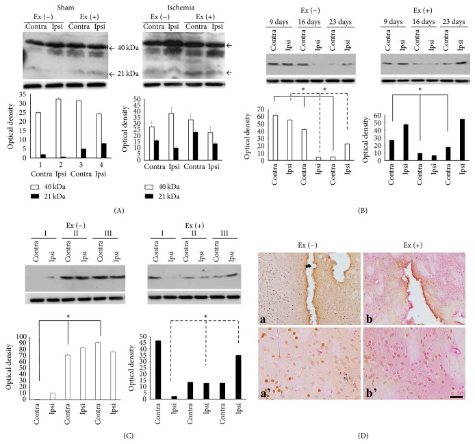
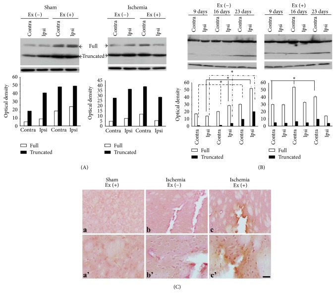
Neurotrophin-3 (NT-3) is a neurotrophic factor that mainly binds to the tyrosine kinase C (trkC) receptor. NT-3 has been shown to have neuroprotective effects in focal cerebral ischemia. Exercise also has ability to induce functional recovery in focal cerebral ischemia. However, the relationship between NT-3, its receptor trkC, and exercise has not been revealed. In this study, we assessed the expressions of NT-3 and trkC in focal cerebral ischemia. We also assessed the expression of NT-3 and trkC with treadmill exercise in focal cerebral ischemia. The results showed that, in a permanent middle cerebral artery occlusion rat model, exercise increased NT-3 and trkC expression. However, the patterns of expression of NT-3 and trkC at different time points varied. These results suggest that exercise-induced functional recovery in focal cerebral ischemia was related to NT-3 and trkC, but the role on times of NT-3 and trkC differed, although trkC is the receptor kinase for NT-3.


Figures

Figure 1
Figure 1
Expression profile of NT-3. (A) There are two forms of NT-3, dimer (40 kDa) and monomer (21 kDa). Exercise in the sham group increased immunoreactivities of the monomer. Exercise in ischemia increased the monomer in the contralateral hemisphere (contra). (B) In the ischemia-nonexercise group, expression of the NT-3 dimer protein decreased from postinfarct day 9 to postinfarct day 23 in the contralateral region. In the ipsilateral region, expression of NT-3 dimer protein was decreased at postinfarct day 9 and then increased at postinfarct day 23. However, in the ischemia-exercise group, expression of the NT-3 dimer protein was decreased at postinfarct day 16 and then increased at postinfarct day 23. (C) The expression of NT-3 dimer protein increased along with severities II and III in the ischemia-nonexercise group. However, expression of NT-3 dimer decreased in the ischemia-exercise group. (D) (a) Immunoreactivity decreased in the core of the infracted region. In contrast, staining increased around the ischemic area. (b) Exercise decreased this contrasting effect. (a') and (b') are magnificent figures of (a) and (b) each. ■ = 25 μm (p < 0.05).
Figure 2
Figure 2
trkC expression profile. (A) There are two forms: full-length (140 kDa) and truncated (90–95 kDa). Ischemia increased both forms, while exercise increased both forms of protein in the bilateral hemispheres, particularly in the contralateral hemisphere (contra) in the ischemic brain. (B) The maximal expression of the two forms was observed at day 23 in the ischemia group. In the exercise group, the full-length form increased with time in the contralateral region, while the expression of the full-length decreased with time in the ipsilateral region. The truncated form was increased at postinfarct day 23 in the contralateral region. (C) (a) In the sham group, exercise itself increased immunoreactivities in both hemispheres, particularly in the vascular structures. (b) Immunoreactivity also increased in the ischemic region. (c) The immunoreactivities were restricted following exercise. (a'), (b'), and (c') are magnificent figures of (a), (b), and (c) each. ■ = 25 μm (p < 0.05).

References

    1. Mozaffarian D., Benjamin E. J., Go A. S., et al. Heart disease and stroke statistics-2015 update : a report from the american heart association. Circulation. 2015;131(4):e29–e39. doi: 10.1161/CIR.0000000000000152. - DOI - PubMed
    1. Wang G., Zhang Z., Ayala C., Dunet D. O., Fang J., George M. G. Costs of hospitalization for stroke patients aged 18–64 years in the United States. Journal of Stroke and Cerebrovascular Diseases. 2014;23(5):861–868. doi: 10.1016/j.jstrokecerebrovasdis.2013.07.017. - DOI - PMC - PubMed
    1. Arya K. N., Pandian S., Verma R., Garg R. K. Movement therapy induced neural reorganization and motor recovery in stroke: a review. Journal of Bodywork and Movement Therapies. 2011;15(4):528–537. doi: 10.1016/j.jbmt.2011.01.023. - DOI - PubMed
    1. Johansson B. B., Zhao L., Mattsson B. Environmental influence on gene expression and recovery from cerebral ischemia. Acta Neurochirurgica Supplement. 1999;73:51–55. - PubMed
    1. Kokaia Z., Zhao Q., Kokaia M., et al. Regulation of brain-derived neurotrophic factor gene expression after transient middle cerebral artery occlusion with and without brain damage. Experimental Neurology. 1995;136(1):73–88. doi: 10.1006/exnr.1995.1085. - DOI - PubMed

MeSH terms

LinkOut - more resources