. 2018 Jul;55(7):5639-5657.
doi: 10.1007/s12035-017-0775-0. Epub 2017 Oct 10.

Physical Exercise Modulates L-DOPA-Regulated Molecular Pathways in the MPTP Mouse Model of Parkinson's Disease

Affiliations

Physical Exercise Modulates L-DOPA-Regulated Molecular Pathways in the MPTP Mouse Model of Parkinson's Disease

Cornelius J H M Klemann et al. Mol Neurobiol. 2018 Jul.

Abstract

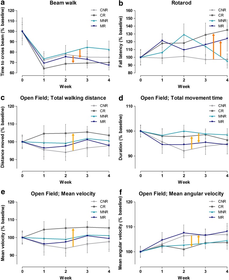
Parkinson's disease (PD) is characterized by the degeneration of dopaminergic (DA) neurons in the substantia nigra pars compacta (SNpc), resulting in motor and non-motor dysfunction. Physical exercise improves these symptoms in PD patients. To explore the molecular mechanisms underlying the beneficial effects of physical exercise, we exposed 1-methyl-4-phenyl-1,2,3,6-tetrahydropyrimidine (MPTP)-treated mice to a four-week physical exercise regimen, and subsequently explored their motor performance and the transcriptome of multiple PD-linked brain areas. MPTP reduced the number of DA neurons in the SNpc, whereas physical exercise improved beam walking, rotarod performance, and motor behavior in the open field. Further, enrichment analyses of the RNA-sequencing data revealed that in the MPTP-treated mice physical exercise predominantly modulated signaling cascades that are regulated by the top upstream regulators L-DOPA, RICTOR, CREB1, or bicuculline/dalfampridine, associated with movement disorders, mitochondrial dysfunction, and epilepsy-related processes. To elucidate the molecular pathways underlying these cascades, we integrated the proteins encoded by the exercise-induced differentially expressed mRNAs for each of the upstream regulators into a molecular landscape, for multiple key brain areas. Most notable was the opposite effect of physical exercise compared to previously reported effects of L-DOPA on the expression of mRNAs in the SN and the ventromedial striatum that are involved in-among other processes-circadian rhythm and signaling involving DA, neuropeptides, and endocannabinoids. Altogether, our findings suggest that physical exercise can improve motor function in PD and may, at the same time, counteract L-DOPA-mediated molecular mechanisms. Further, we hypothesize that physical exercise has the potential to improve non-motor symptoms of PD, some of which may be the result of (chronic) L-DOPA use.

Keywords: (Non-)motor function; L-DOPA; MPTP; Molecular landscape; Parkinson’s disease; Physical exercise.


Conflict of interest statement

Conflict of Interest

The authors declare that they have no conflict of interest.

Figures

Fig. 1
Fig. 1
Effect of MPTP. Results of the behavioral tests in week 0 (set as the baseline (100%) for the effect of physical exercise, which is shown in Fig. 2). No effect of MPTP was shown for the beam walk (a), or rotarod (b) tests, but MPTP significantly affects the parameters in the open field (cf). **p < 0.01; ***p < 0.005, mean + SEM, n = 28 for controls (saline-treated), and n = 23 for MPTP-treated mice
Fig. 2
Fig. 2
Effect of physical exercise and interaction with MPTP compared to the baseline (week 0); results of the behavioral tests in week 0–4. Measurements in week 1–4 were normalized to week 0 (100%). Dark-orange arrows indicate the main effect of physical exercise (a, b), and light orange arrows show the effect of the interaction between physical exercise and MPTP treatment (cf) mean ± SEM n = 14 for both CNR and CR, n = 10 for MNR and n = 13 for MR. CNR, control not running; CR control running, MNR MPTP-treated but not running, MR MPTP-treated and running
Fig. 3
Fig. 3
TH+ neurons in the SNpc and VTA. The upper panel shows a representative picture for each of the four treatment groups and the lower panel shows the average number of TH+ neurons in the SNpc and the VTA per treatment group. *p < 0.05, mean + SEM, n = 5 for CNR and MR and n = 4 for CR, and MNR for both brain areas. CNR control not running, CR control running, MNR MPTP-treated but not running, MR MPTP-treated and running, SNpc substantia nigra pars compacta, VTA ventral tegmental area
Fig. 4
Fig. 4
Landscape of proteins encoded by the mRNAs regulated by physical exercise and the upstream regulator L-DOPA in the SN. mRNAs differentially expressed in the SN due to physical exercise in MPTP-treated mice are shown in gray. Blue proteins are additional genes/proteins that are associated with PD through genetic and/or expression studies, whereas white proteins have no known link with PD. The direction of effect of physical exercise (measured) and L-DOPA (from literature) on the expression of these mRNAs is depicted through colored borders. L-DOPA-activated proteins are shown with purple writing for the protein name, and familial PD proteins are shown with a blue border
Fig. 5
Fig. 5
Landscape of proteins encoded by the mRNAs regulated by physical exercise and the upstream regulator L-DOPA in the VM. mRNAs differentially expressed in the VM due to physical exercise in MPTP-treated mice are shown in gray. Blue proteins are additional genes/proteins that are associated with PD through genetic and/or expression studies, whereas white proteins have no known link with PD. The direction of effect of physical exercise (measured) and L-DOPA (from literature) on the expression of these mRNAs is depicted through colored borders. L-DOPA-activated proteins are shown with purple writing for the protein name, and familial PD proteins are shown with a blue border
Fig. 6
Fig. 6
Overview of the brain areas analyzed, and the top upstream regulators, and processes per area. The brain areas are shown in a simplified model of the basal ganglia circuitry. Green, red, and gray triangles depict positive (> 2), negative (< −2) or non-significant z scores, respectively, from the enrichment analyses of the physical exercise-regulated mRNAs in MPTP-treated mice. DL dorsolateral striatum, GPe globus pallidus external, GPi globus pallidus internal, PFC prefrontal cortex, PPN pedunculopontine nucleus, SNpc substantia nigra pars compacta, SNr substantia nigra reticularis, STN subthalamic nucleus, VM ventromedial striatum, VTA ventral tegmental area

Similar articles

Cited by

References

    1. Goetz CG, Tilley BC, Shaftman SR, Stebbins GT, Fahn S, Martinez-Martin P, et al. Movement Disorder Society-sponsored revision of the Unified Parkinson's Disease Rating Scale (MDS-UPDRS): scale presentation and clinimetric testing results. Mov Disord. 2008;23(15):2129–2170. - PubMed
    1. David FJ, Robichaud JA, Leurgans SE, Poon C, Kohrt WM, Goldman JG, et al. Exercise improves cognition in Parkinson's disease: The PRET-PD randomized, clinical trial. Mov Disord. 2015;30(12):1657–1663. - PMC - PubMed
    1. Sveinbjornsdottir S. The clinical symptoms of Parkinson's disease. J Neurochem. 2016;139(Suppl 1):318–324. - PubMed
    1. Cotzias GC, Van Woert MH, Schiffer LM. Aromatic amino acids and modification of parkinsonism. N Engl J Med. 1967;276(7):374–379. - PubMed
    1. Fox SH, Katzenschlager R, Lim SY, Ravina B, Seppi K, Coelho M, et al. The Movement Disorder Society evidence-based medicine review update: treatments for the motor symptoms of Parkinson's disease. Mov Disord. 2011;26(Suppl 3):S2–41. - PubMed

MeSH terms