Review
. 2017 Oct 30;46(21):6638-6663.
doi: 10.1039/c7cs00521k.

Self-assembling prodrugs

Affiliations
Review

Self-assembling prodrugs

Andrew G Cheetham et al. Chem Soc Rev. .

Abstract

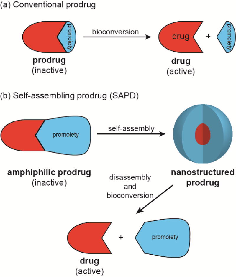
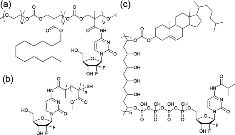
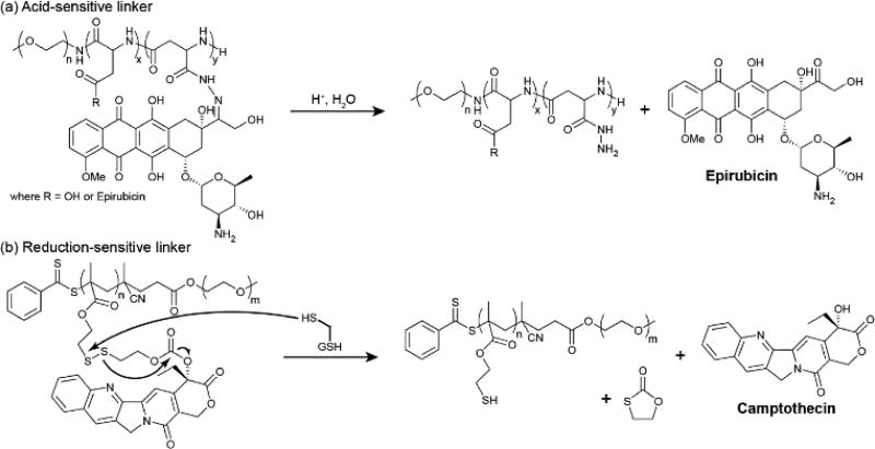
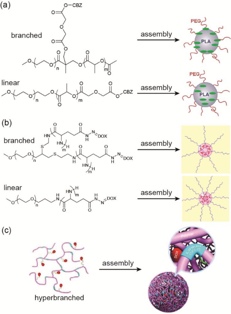
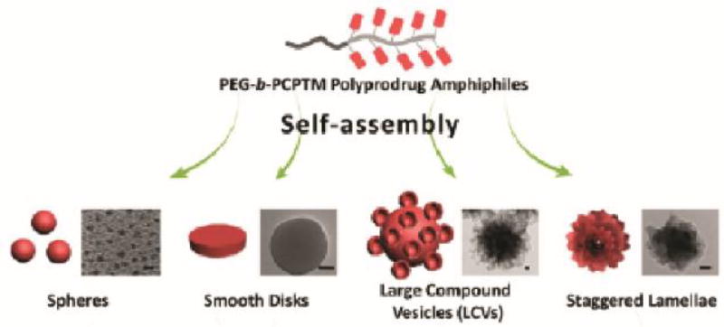
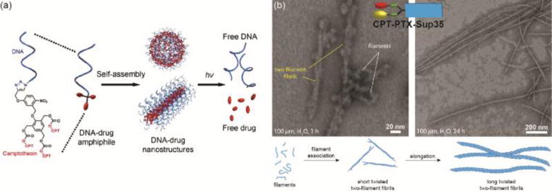
Covalent modification of therapeutic compounds is a clinically proven strategy to devise prodrugs with enhanced treatment efficacies. This prodrug strategy relies on the modified drugs that possess advantageous pharmacokinetic properties and administration routes over their parent drug. Self-assembling prodrugs represent an emerging class of therapeutic agents capable of spontaneously associating into well-defined supramolecular nanostructures in aqueous solutions. The self-assembly of prodrugs expands the functional space of conventional prodrug design, affording a possible pathway to more effective therapies as the assembled nanostructure possesses distinct physicochemical properties and interaction potentials that can be tailored to specific administration routes and disease treatment. In this review, we will discuss the various types of self-assembling prodrugs in development, providing an overview of the methods used to control their structure and function and, ultimately, our perspective on their current and future potential.


Figures

Fig. 1
Fig. 1
Illustration of (a) conventional prodrugs and (b) self-assembling prodrugs.
Fig. 2
Fig. 2
Kataoka’s initial design of a self-assembling polymer–conjugate, PEG-b-P(Asp(DOX)).
Fig. 3
Fig. 3
Examples of self-assembling PDCs using PEG-b-P(Glu) as the polymer backbone.
Fig. 4
Fig. 4
Examples of self-assembling PDCs synthesized using alternative polymer backbones.
Fig. 5
Fig. 5
Examples of hydrophilic drug containing PDCs.
Fig. 6
Fig. 6
Examples of stimuli-sensitive linkers and their release mechanisms. (a) Acid-sensitive hydrazone linkers are cleaved under the acidic conditions found in the lysosomal and endosomal cellular compartments. (b) Reduction-sensitive disulphide linkers are cleaved in the presence of intracellular reductants such as glutathione (GSH) or cysteine.
Fig. 7
Fig. 7
Variations in the polymer architecture can affect the drug’s position and release characteristics. Panel (a) was adapted from reference [86] with permission from the American Chemical Society (ACS), copyright 2013. Panel (b) was adapted from reference [87] with permission from the Multidisciplinary Digital Publishing Institute (MDPI), copyright 2014. Panel (c) was adapted from reference [88] with permission from ACS, copyright, 2016.
Fig. 8
Fig. 8
Varying experimental parameters to influence assembly during nanoprecipitation of a PDC. Reproduced from reference [91] with permission from ACS, copyright 2013.
Fig. 9
Fig. 9
Strategies for combination therapy using self-assembling PDCs.
Fig. 10
Fig. 10
Biopolymer–drug conjugates. (a) Chilkoti’s DOX-containing polypeptide–drug conjugate based on recombinant elastin-like peptides. (b) A Cellax-based carbohydrate–drug conjugate synthesized by Ernsting et al.
Fig. 11
Fig. 11
Chemical Structure of 4-(N)-trisnorsqualenoylgemcitabine (SQdFdC). After dropwise addition of the drug solution into a large body of water, successive removal of solvent will form stabilized nanoparticles. Transmission electron micrograph (TEM) of the resulting SQdFdC nanoassemblies is shown (scale bar 50nm). The TEM image was adapted from reference [128] with permission from ACS, copyright 2006.
Fig. 12
Fig. 12
Chemical structures of Shen’s diCPT-OEG and CPT-OEG prodrugs. These drug conjugates were capable of forming nanovesicles with the ability to encapsulate DOX (purple spheres). Figure was reproduced from reference [145] with permission from ACS, copyright 2010.
Fig. 13
Fig. 13
Examples of drug–drug conjugates in which two drugs are linked together, either directly or through a short linker.
Fig. 14
Fig. 14
Enzyme-instructed self-assembly (EISA) strategy developed by Xu and co-workers. After enzyme-activated dephosphorylation, the taxol conjugates spontaneously self-assemble into nanofibers (A). The chemical structure of the corresponding taxol conjugate (B). A dense network of nanofibers contribute to the formation of a hydrogel (C, D). Adapted from reference [158] with permission from ACS, copyright 2009.
Fig. 15
Fig. 15
An illustration of the EISA mechanism in the pericellular space, where a chemical modification helps trigger the self-assembly of NDP1 into nanofibrils. (A) Dephosphorylation, by alkaline phosphatase (ALP), removes the phosphate group and in turn activates the NDP1 precursor. (B) The molecular structures of pNDP1 and NDP1 where the orange segment is the fluorophore NBD and red indicates the phosphate enzymatic trigger. (C) The overexpression of ALP by cancer cells limits dephosphorylation proximal to the cell membrane. This stimulates apoptosis. (D) Image of the fluorescent fibrils selectively forming around the HeLa cancer cells (white arrows) in a co-culture with Hs-5 stroma cells (white arrowheads). With time, the amount of self-assembled fibrils increases with the majority focused around HeLa cells. The left image was at time 0 h, middle at 6 h, and right at 24 h. Scale bar represents 10 µm. Adapted from reference [166] with permission from Elsevier, copyright 2016.
Fig. 16
Fig. 16
Illustration of a CPT-based drug amphiphile that can spontaneously self-assemble into nanofibers upon ageing in water. Adapted from reference [183] with permission from ACS, copyright 2013.
Fig. 17
Fig. 17
Morphological changes induced by structural alterations. (a) Zhang’s nucleic acid–drug conjugates formed spherical or cylindrical nanostructures depending on the number of DNA base pairs in the molecular structure. (b) Cui and co-workers showed that the incorporation of two structurally different drug molecules, CPT and PTX in this case, can significantly alter the self-assembly pathway and the emergent nanostructure. Panel (a) is reproduced from reference [198] with permission from ACS, copyright 2015. Panel (b) is adapted from reference [200] with permission from the Royal Society of Chemistry (RSC), copyright 2014.

References

    1. Albert A. Nature. 1958;182:421–423. - PubMed
    1. Rautio J, Kumpulainen H, Heimbach T, Oliyai R, Oh D, Jarvinen T, Savolainen J. Nature Reviews Drug Discovery. 2008;7:255–270. - PubMed
    1. Kevin B, Robert W, Iain G, Kevin D. Current Drug Metabolism. 2003;4:461–485. - PubMed
    1. Stella VJ, Nti-Addae KW. Advanced Drug Delivery Reviews. 2007;59:677–694. - PubMed
    1. Simplício LA, Clancy MJ, Gilmer FJ. Molecules. 2008;13 - PMC - PubMed