. 2017 Oct 11;12(10):e0186194.
doi: 10.1371/journal.pone.0186194. eCollection 2017.

Diacetylenic lipids in the design of stable lipopolymers able to complex and protect plasmid DNA

Affiliations

Diacetylenic lipids in the design of stable lipopolymers able to complex and protect plasmid DNA

C Facundo Temprana et al. PLoS One. .

Abstract

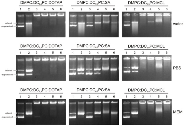
Different viral and non-viral vectors have been designed to allow the delivery of nucleic acids in gene therapy. In general, non-viral vectors have been associated with increased safety for in vivo use; however, issues regarding their efficacy, toxicity and stability continue to drive further research. Thus, the aim of this study was to evaluate the potential use of the polymerizable diacetylenic lipid 1,2-bis(10,12-tricosadiynoyl)-sn-glycero-3-phosphocholine (DC8,9PC) as a strategy to formulate stable cationic lipopolymers in the delivery and protection of plasmid DNA. Cationic lipopolymers were prepared following two different methodologies by using DC8,9PC, 1,2-dimyristoyl-sn-glycero-3-phosphocholine (DMPC), and the cationic lipids (CL) 1,2-dioleoyl-3-trimethylammonium-propane (DOTAP), stearylamine (SA), and myristoylcholine chloride (MCL), in a molar ratio of 1:1:0.2 (DMPC:DC8,9PC:CL). The copolymerization methodology allowed obtaining cationic lipopolymers which were smaller in size than those obtained by the cationic addition methodology although both techniques presented high size stability over a 166-day incubation period at 4°C. Cationic lipopolymers containing DOTAP or MCL were more efficient in complexing DNA than those containing SA. Moreover, lipopolymers containing DOTAP were found to form highly stable complexes with DNA, able to resist serum DNAses degradation. Furthermore, neither of the cationic lipopolymers (with or without DNA) induced red blood cell hemolysis, although metabolic activity determined on the L-929 and Vero cell lines was found to be dependent on the cell line, the formulation and the presence of DNA. The high stability and DNA protection capacity as well as the reduced toxicity determined for the cationic lipopolymer containing DOTAP highlight the potential advantage of using lipopolymers when designing novel non-viral carrier systems for use in in vivo gene therapy. Thus, this work represents the first steps toward developing a cationic lipopolymer-based gene delivery system using polymerizable and cationic lipids.


Conflict of interest statement

Competing Interests: The authors have declared that no competing interests exist.

Figures

Fig 1
Fig 1. Polymerization confirmation.
Absorbance as a function of wavelength (nm) for the DMPC:DC8,9PC:DOTAP (1:1:0.2), DMPC:DC8,9PC:MCL (1:1:0.2) and DMPC:DC8,9PC:SA (1:1:0.2) mixtures, prepared with the copolymerization methodology, after 20 UV irradiation cycles. Peaks observed around 480 and 520 nm are indicative of polymer formation.
Fig 2
Fig 2. Study of cationic lipopolymer/DNA interaction.
Gel retardation assay for (a) DMPC:DC8,9PC:DOTAP (1:1:0.2), DMPC:DC8,9PC:SA (1:1:0.2), DMPC:DC8,9PC:MCL (1:1:0.2) and DMPC:DC8,9PC (1:1) mixtures incubated at 0:1, 6:1, 8:1, 10:1, 12:1, 14:1, 16:1 cationic lipopolymer or lipopolymer/pCH110 plasmid DNA ratios (mol of lipids: mol of base pairs) and (b) DMPC:DC8,9PC:DOTAP (1:1:0.2), DMPC:DC8,9PC:SA (1:1:0.2), DMPC:DC8,9PC:MCL (1:1:0.2) and DMPC:DC8,9PC (1:1) mixtures incubated at 0:1, 8:1, 16:1, 24:1, 30:1, 36:1, 42:1 cationic lipopolymer or lipopolymer/pDsRed plasmid DNA ratios (mol of lipids: mol of base pairs). All lanes were loaded with 1 μg of plasmid DNA.
Fig 3
Fig 3. Stoichiometry of the cationic lipopolymer/DNA complex.
Percentage of plasmid DNA association as a function of cationic lipopolymer/pCH110 (a and b) or pDsRed (c and d) plasmid DNA ratios (mol of lipids: mol of base pairs) for the DMPC:DC8,9PC:DOTAP (1:1:0.2), DMPC:DC8,9PC:SA (1:1:0.2), and DMPC:DC8,9PC:MCL (1:1:0.2) formulations.
Fig 4
Fig 4. Effect of different incubation media on the cationic lipopolymer/DNA interaction.
The different cationic lipopolymers were incubated with the pDsRed plasmid DNA in a 16:1 (mol of lipids: mol of base pairs) ratio in the medium (water, PBS or MEM) indicated on the right. Each lane was loaded with 1 μg of plasmid DNA. Lanes correspond to: (1) pDsRed alone with an additional 10-min incubation in the indicated medium, (2) pDsRed alone with an additional 10-min incubation in the presence of 10% v/v FBS, (3) cationic lipopolymer (stated above the gel picture)/pDsRed plasmid DNA complex formed in the indicated medium with an additional 10-min incubation without FBS, (4) cationic lipopolymer (stated above the gel picture)/pDsRed plasmid DNA complex formed in the indicated medium with an additional 10-min incubation in the presence of 10% v/v FBS, (5) cationic lipopolymer (stated above the gel picture)/pDsRed plasmid DNA complex formed in the indicated medium with an additional 10-min incubation in the presence of 50% v/v FBS, (6) cationic lipopolymer (stated above the gel picture)/pDsRed plasmid DNA complex formed in the indicated medium with 10% v/v FBS and with an additional 10-min incubation in the presence of 50% v/v FBS. The two main topological plasmid conformations, relaxed and negatively supercoiled, are indicated with arrows on the left of the figure noted as relax and -supercoiled, respectively.
Fig 5
Fig 5. Serum nucleases digestion assay.
The different cationic lipopolymers DMPC:DC8,9PC:DOTAP (1:1:0.2) and DMPC:DC8,9PC:MCL (1:1:0.2)/pDsRed plasmid DNA complexes (16:1 mol of lipids: mol of base pairs ratio) were formed in water, PBS or MEM (indicated on the right). Each lane was loaded with 1 μg of plasmid DNA. Lanes correspond to: (1) pDsRed alone with a 24-h incubation in the indicated medium, (2) pDsRed alone with a 24-h incubation in the presence of 50% v/v FBS, (3) cationic lipopolymer (stated above the gel picture)/pDsRed plasmid DNA complex formed in the indicated medium with a 24-h incubation in the indicated medium without FBS, and (4) cationic lipopolymer (stated above the gel picture)/pDsRed plasmid DNA complex formed in the indicated medium with a 24-h incubation in the presence of 50% v/v FBS. The two main topological plasmid conformations, relaxed and negatively supercoiled, are indicated with arrows on the left of the figure noted as relax and -supercoiled, respectively. Degraded DNA is also indicated with an arrow on the left of the figure.
Fig 6
Fig 6. Optimization of the study of cationic lipopolymer/DNA interaction by flow cytometry.
Flow cytometry analysis results for non-polymerized DMPC:DC8,9PC:MCL (1:1:0.2) liposomes, used to set control values of (a) SSC-H values (related to particle complexity) versus FSC-H values (related to particle size) and (b) FL1 values (where SYBR® Green I-labeled plasmid DNA fluorescence is detected) and FL2 values (where cationic lipopolymer fluorescence is detected). (c) and (d) show the results for polymerized DMPC:DC8,9PC:MCL (1:1:0.2) for SSC-H versus FSC-H and FL2 versus FL1 values respectively. (e) and (f) show the results for non-polymerized DMPC:DC8,9PC:MCL (1:1:0.2)/SYBR® Green I-labeled pDsRed plasmid DNA complexes for SSC-H and FSC-H values and FL2 versus FL1 values respectively.
Fig 7
Fig 7. Study of cationic lipopolymer/DNA interaction by flow cytometry.
Flow cytometry analysis results for (a) SSC-H versus FSC-H and (b) FL2 versus FL1 values for polymerized DMPC:DC8,9PC:DOTAP (1:1:0.2)/SYBR® Green I-labeled pDsRed plasmid DNA complexes and (c) SSC-H versus FSC-H and (d) FL2 versus FL1 values for polymerized DMPC:DC8,9PC:MCL (1:1:0.2)/SYBR® Green I-labeled pDsRed plasmid DNA complexes.
Fig 8
Fig 8. Study of cationic lipopolymer/DNA interaction by flow cytometry.
Flow cytometry analysis results for (a) SSC-H versus FSC-H and (b) FL2 versus FL1 values for polymerized DMPC:DC8,9PC:DOTAP (1:1:0.2)/SYBR® Green I-labeled pDsRed plasmid DNA complexes. Different regions were drawn based on SSC-H and FSC-H values (a) and the fluorescence values for FL1 and FL2 corresponding to each region are marked in the FL2 versus FL1 graph (b) with the same borderline shown by the same style line arrow.
Fig 9
Fig 9. Cytotoxicity determination.
Cell viability percentage as a function of total lipid concentration (mM) for L929 cells incubated with cationic lipopolymers alone (a) or complexed with pDsRed plasmid DNA in a 16:1 (mol of lipids: mol of base pairs) ratio (b); and for Vero cells incubated with cationic lipopolymers alone (c) or complexed with pDsRed plasmid DNA in a 16:1 (mol of lipids: mol of base pairs) ratio (d). Statistical analysis was performed by Two-Way ANOVA with Tukey´s multiple comparisons post-test. *<0.05; **<0.01; ***<0.001; ****<0.0001.

Similar articles

Cited by

References

    1. He C-X, Tabata Y, Gao J-Q. Non-viral gene delivery carrier and its three-dimensional transfection system. International journal of pharmaceutics. 2010;386(1):232–42. - PubMed
    1. Morille M, Passirani C, Vonarbourg A, Clavreul A, Benoit J-P. Progress in developing cationic vectors for non-viral systemic gene therapy against cancer. Biomaterials. 2008;29(24):3477–96. - PubMed
    1. Naldini L. Gene therapy returns to centre stage. Nature. 2015;526(7573):351–60. doi: 10.1038/nature15818 - DOI - PubMed
    1. Oliveira A, Ferraz M, Monteiro F, Simoes S. Cationic liposome—DNA complexes as gene delivery vectors: development and behaviour towards bone-like cells. Acta Biomaterialia. 2009;5(6):2142–51. doi: 10.1016/j.actbio.2009.02.019 - DOI - PubMed
    1. Porras G, Bezard E. Preclinical development of gene therapy for Parkinson's disease. Experimental neurology. 2008;209(1):72–81. doi: 10.1016/j.expneurol.2007.08.003 - DOI - PubMed

MeSH terms

LinkOut - more resources