. 2017 Oct 11;16(1):157.
doi: 10.1186/s12943-017-0724-6.

SUMO1 modification of KHSRP regulates tumorigenesis by preventing the TL-G-Rich miRNA biogenesis

Affiliations

SUMO1 modification of KHSRP regulates tumorigenesis by preventing the TL-G-Rich miRNA biogenesis

Haihua Yuan et al. Mol Cancer. .

Abstract

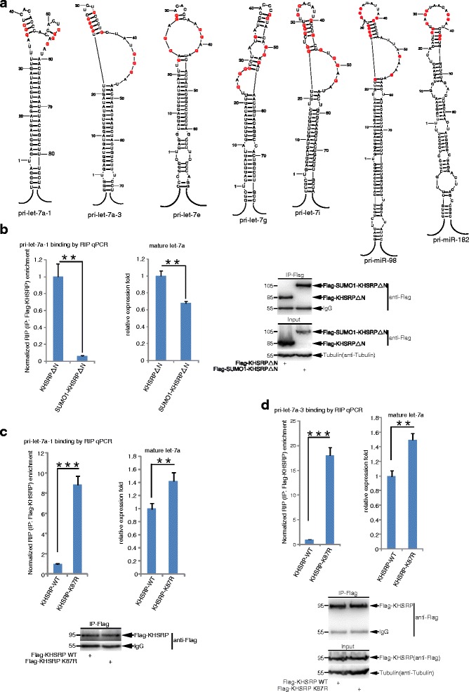
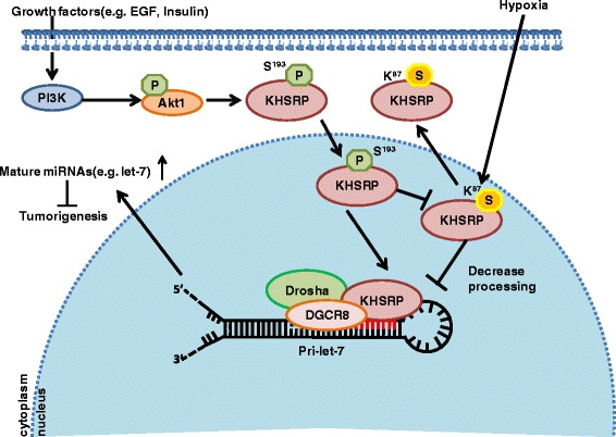
Background: MicroRNAs (miRNAs) are important regulators involved in diverse physiological and pathological processes including cancer. SUMO (small ubiquitin-like modifier) is a reversible protein modifier. We recently found that SUMOylation of TARBP2 and DGCR8 is involved in the regulation of the miRNA pathway. KHSRP is a single stranded nucleic acid binding protein with roles in transcription and mRNA decay, and it is also a component of the Drosha-DGCR8 complex promoting the miRNA biogenesis.

Methods: The in vivo SUMOylation assay using the Ni2+-NTA affinity pulldown or immunoprecipitation (IP) and the in vitro E.coli-based SUMOylation assay were used to analyze SUMOylation of KHSRP. Nuclear/Cytosol fractionation assay and immunofluorescent staining were used to observe the localization of KHSRP. High-throughput miRNA sequencing, quantantive RT-PCR and RNA immunoprecipitation assay (RIP) were employed to determine the effects of KHSRP SUMO1 modification on the miRNA biogenesis. The soft-agar colony formation, migration, vasculogenic mimicry (VM) and three-dimensional (3D) cell culture assays were performed to detect the phenotypes of tumor cells in vitro, and the xenograft tumor model in mice was conducted to verify that SUMO1 modification of KHSRP regulated tumorigenesis in vivo.

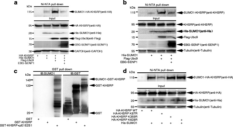
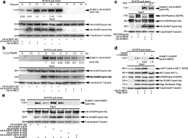
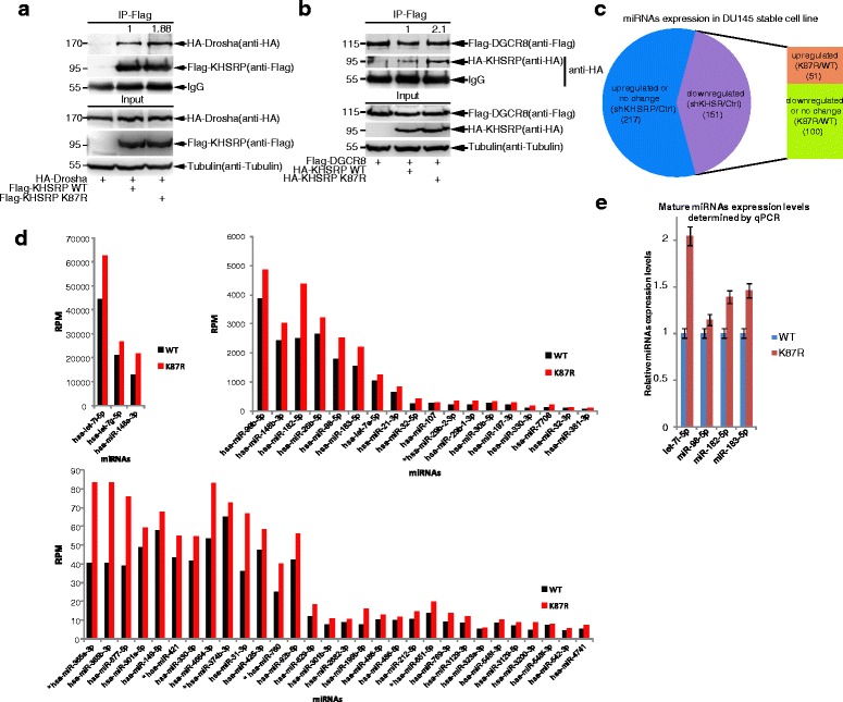
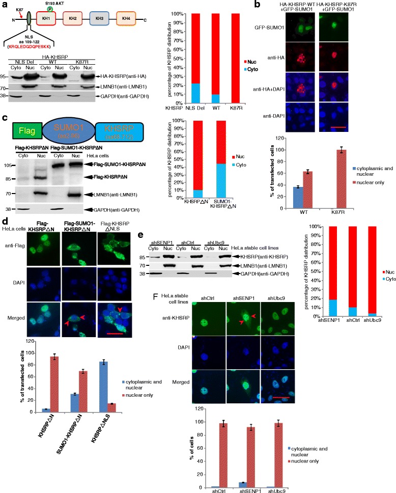
Results: KHSRP is modified by SUMO1 at the major site K87, and this modification can be increased upon the microenvironmental hypoxia while reduced by the treatment with growth factors. SUMO1 modification of KHSRP inhibits its interaction with the pri-miRNA/Drosha-DGCR8 complex and probably increases its translocation from the nucleus to the cytoplasm. Consequently, SUMO1 modification of KHSRP impairs the processing step of pre-miRNAs from pri-miRNAs which especially harbor short G-rich stretches in their terminal loops (TL), resulting in the downregulation of a subset of TL-G-Rich miRNAs such as let-7 family and consequential tumorigenesis.

Conclusions: Our data demonstrate how the miRNA biogenesis pathway is connected to tumorigenesis and cancer progression through the reversible SUMO1 modification of KHSRP.

Keywords: KHSRP; SUMO1 modification; TL-G-Rich miRNA biogenesis; Tumorgenesis.


Conflict of interest statement

Ethics approval

All animal studies were conducted with the approval and guidance of Shanghai Jiao Tong University Medical Animal Ethics Committees.

Consent for publication

All authors read and approved the final manuscript, and consent to publish.

Competing interests

The authors declare that they have no competing interests.

Figures

Fig. 1
Fig. 1
KHSRP is modified by SUMO1 at the major site K87 in vitro and in cells. a-b Exogenous and endogenous KHSRP in cells are modified by SUMO1. 293T cells transfected with indicated plasmids were lysed and pulled down with Ni2+-NTA resin for SUMOylation assay, and SUMO1 modification of KHSRP was analyzed by Western blotting with indicated antibodies. c SUMO1 modification of KHSRP is verified by in vitro E.coli-based SUMOylation assay. Plasmid pGEX-4T-1-KHSRP was co-transformed with or without pE1E2SUMO1 plasmid into E.coli BL21 (DE3). After GST pull-down purification, Western blotting was conducted with anti-SUMO1 antibody and the same membrane was detected with anti-GST antibody after stripping. d Mutation of K87R weakens SUMO1 modification of KHSRP in 293T cells. The construct pEF-5HA-KHSRP-WT, or -K87R, or -K359R, or -K628R was co-transfected with His-SUMO1 into 293T cells. 48 h after transfection, cells were lysed for the SUMOylation assay with Ni2+-NTA resin
Fig. 2
Fig. 2
SUMO1 modification of KHSRP is regulated by hypoxia, hydrogen peroxide and growth factors. a Hypoxia upregulates SUMO1 modification of KHSRP. 293T cells transfected with His-SUMO1 and HA-KHSRP-WT or HA-KHSRP-K87R were cultured in 1% oxygen condition (hypoxia) for 6, 12 h before cells were harvested. Ni2+-NTA resin pull down was performed to detect the SUMO1 modification of exogenous KHSRP. b Hydrogen peroxide (H2O2) downregulates SUMO1 modification of KHSRP. 293T cells transfected with His-SUMO1 and HA-KHSRP-WT or HA-KHSRP-K87R were treated with 100 μM of H2O2 for 1.5, 3 h before cells were harvested. Ni2+-NTA resin pull down was performed to detect the SUMO1 modification of KHSRP. c EGF downregulates SUMO1 modification of KHSRP. 36 h after transfection with His-SUMO1 and HA-KHSRP-WT, 293T cells were starved overnight and then stimulated with EGF (50 ng/ml) for 5 min before lysed for Ni2+-NTA pull down, and followed by Western blotting with indicated antibodies. d Insulin downregulates SUMO1 modification of KHSRP. 293T cells transfected with His-SUMO1 and Flag-Ubc9 were treated with insulin (1 μM) for 1 h or LY294002 (25 μM) for 16 h before cells were harvested. Ni2+-NTA resin pull down was performed to detect the SUMO1 modification of endogenous KHSRP. p-AKT1 (S473) antibody was used to detect the phosphorylation level of AKT1. e Phosphorylation of KHSRP downregulates SUMO1 modifcation of KHSRP. 293T cells transfected with His-SUMO1 and HA-KHSRP-WT, −K87R, −S193A, −K87R-S193A, −S193D, or -K87R-S193D were lysed for the SUMOylation assay with the Ni2+-NTA resin pull down. The band intensities were calculated by ImageJ software and the ratios were quantified (a-d)
Fig. 3
Fig. 3
SUMOylation of KHSRP is involved in tumorigenesis. a KHSRP-K87R downregulates the anchorage-independent growth in DU145 stable cell lines. In soft agar colony forming assays, stable cell lines DU145 shRNA control, shKHSRP, shKHSRP-KHSRP-WT or shKHSRP-KHSRP-K87R were seeded in 2 ml of medium containing 5% FBS with 0.35% agar at 2 × 103 cells/well and layered onto the base. The photographs were taken 21 days later and the number of colonies was scored. b KHSRP-K87R downregulates the migration ability in DU145 stable cell lines. The RTCA migration assay was performed to detect the migration ability in above stable DU145 cell lines with xCELLigene RTCA-DP instrument. The kinetic cell index of their migration was recorded every 15 min for 24 h (left panel) and the relative slope value was calculated (right panel). c KHSRP-K87R downregulates the invasive ability in above stable DU145 cell lines. The 3D–culture assay was performed to detect the invasive ability of DU145 stable cell lines. The photos were taken at day 7. The first image was taken under the white light, and the green signals indicates the expression of GFP (Green Fluorescent Protein) in the plasmid CD513B-HA-KHSRP. d KHSRP-K87R downregulates the aggressive ability in DU145 stable cell lines in vasculogenic mimicry (VM) assay. VM assay was performed to detect the aggressive ability in above stable DU145 cell lines. The photos were taken 20 h later. Scale: 500 μm. Independent experiments (a-d) were repeated three times. e KHSRP-K87R suppresses xenograft tumor growth in vivo. 5 male BALB/c nude mice were injected subcutaneously with stable DU145 cell lines (2.5 × 106 cells/each) expressing the shRNA control in the left back and shKHSRP in the right back, respectively. Another 5 male BALB/c nude mice were injected subcutaneously with stable DU145 cell line expressing shKHSRP-KHSRP-WT in the left back and shKHSRP-KHSRP-K87R in the right back, respectively. Mice were sacrificed 5 weeks later, and tumors were dissected (upper panel) and assessed by weight (low panel). (a-d) f KHSRP SUMOylation could be detected in tumors of nude mice. The tumors of nude mice, which was chosen from the groups of shKHSRP, shKHSRP-KHSRP-WT or -K87R, were lysed in NEM-RIPA as described in the Methods. The proteins was immunoprecipitated by anti-SUMO1 antibody, Western blotting was detected with anti-HA antibody
Fig. 4
Fig. 4
SUMOylation of KHSRP at K87 inhibits the biogenesis of miRNAs. (A-B) The mutation K87R of KHSRP increases its interaction with Drosha and DGCR8. 293T cells were transfected with HA-Drosha and Flag-KHSRP-WT or Flag-KHSRP-K87R (a), or transfected with Flag-DGCR8 and HA-KHSRP-WT or HA-KHSRP -K87R (b). 48 h after transfection, cells were lysed for immunoprecipitation with anti-Flag antibody, and followed by Western blotting with indicated antibodies. The band intensities were calculated by ImageJ software and the ratios were quantified. c KHSRP SUMOylation regulates the biogenesis of a subset of miRNAs. High-throughput deep sequencing data showed that there were 368 miRNAs whose RPM (reads per million) > 10 RPM in all expressed miRNAs of DU145 control cell line. 151 of these miRNAs whose expression in DU145-shKHSRP cells were downregulated compared to those in DU145-shRNA control cells. 51 of 151 miRNAs whose expression were upregulated in DU145 shKHSRP-KHSRP-K87R cells compared to those in DU145 shKHSRP-KHSRP-WT cells. d KHSRP-K87R promotes some miRNAs biogenesis. According to sequencing data, a total of 51 miRNAs whose expression were upregulated in DU145 shKHSRP-KHSRP-K87R cells compared to those in DU145 shKHSRP-KHSRP-WT cells. Among 51 miRNAs, there were only 6 miRNAs which harbor only one single G or none G (labeled with *). Detailed can be seen Additional file 10: Table S4. e The biogenesis of some miRNAs promoted by KHSRP-K87R was validated by qRT-PCR. qRT-PCR was performed to assess the expression of endogenous let-7i-5p, miR-98-5p, miR-182a-5p and miR-183a-5p in DU145 shKHSRP-KHSRP-WT and shKHSRP-KHSRP-K87R stable cell lines
Fig. 5
Fig. 5
SUMO1 modification of KHSRP promotes its cytoplasmic localization. a K87R mutation affects the localization of HA-KHSRP. HeLa cells were transfected with HA-KHSRP-WT, HA-KHSRP-K87R or HA-KHSRP-ΔNLS, respectively. Nuclear and cytosolic fractions were extracted by the Nuclear/Cytosol fractionation kit. Nuclear and cytoplasmic fraction extracts were immunoblotted with indicated antibodies. b SUMO1 modification affects the localization of HA-KHSRP by using the method of immunofluorescent staining. HeLa cells transfected with HA-KHSRP-WT or HA-KHSRP-K87R with GFP-SUMO1 were stained with the primary antibody anti-HA (Rabbit), and then with the second antibody of Alexa Fluor 568 anti-rabbit. DAPI staining was to visualize the nucleus. The green signals indicated the expression of GFP-SUMO1 carrying Green Fluorescent Protein and the images of GFP-SUMO1 were directly taken without staining. All the images were taken by Nikon microscope. Scale bar, 25 μm. c-d SUMO1 modification of KHSRP promotes its cytosolic localization. Flag-SUMO1-KHSRPΔN was constructed by replacing the N-terminal (aa 1–67) of KHSRP with Flag-tagged SUMO1(aa 2–96). c HeLa cells transfected with Flag-KHSRPΔN or Flag-SUMO1-KHSRPΔN were extracted by the Nuclear/Cytosol fractionation kit, and followed by immunoblotting with indicated antibodies. d HeLa cells transfected with Flag-KHSRPΔN, Flag-SUMO1-KHSRPΔN or Flag-KHSRPΔNLS were stained using the primary antibody of anti-Flag M2 and the secondary antibody of Alexa Fluor 488 (anti-mouse). e-f SUMOylation increases the cytosolic localization of endogenous KHSRP. e HeLa-shControl, HeLa-shSENP1 and HeLa-shUbc9 cells were extracted by the Nuclear/Cytosol fractionation kit, then immunoblotted with indicated antibodies. f These three cell lines were stained using the primary antibody anti-KHSRP (Rabbit) and the secondary antibody Alexa Fluor 488 (anti-rabbit). Images were taken by Nikon microscope, and the cytoplasmic location of KHSRP was indicated by red arrows. The same scale bar (25 μm) was used in all images (b, d, f). The staining cells numbers were counted with the Image J software and the statistical analysis was performed (b, d, f). The band intensities were calculated by Image J and quantified by normalization to GAPDH and LMNB1, the percentage of nuclear/cytosolic fraction of every sample was calculated (a, c, e)
Fig. 6
Fig. 6
SUMO1 modification of KHSRP interferes its interaction with pri-miRNAs. a Pri-miRNAs whose mature miRNAs were upregulated by KHSRP-K87R were analyzed by using the RNAstructure software. All of pri-let-7e, pri-let-7g, pri-let-7i, pri-miR-98 and pri-miR-182 harbored G-rich stretches in their terminal loops, as like pri-let-7a-1 and pri-let-7a-3. b KHSRPΔN fusing with SUMO1 decreases its interaction with pri-let-7a-1 and the mature let-7a production. 293T cells transfected CD513B-pri-let-7a-1 and Flag-KHSRPΔN, or Flag-SUMO1-KHSRPΔN were lysed for RIP with anti-Flag antibody, then treated with Trizol, and followed by qRT-PCR for pri-let-7a-1. The relative recruitment fold of pri-let-7a-1 by KHSRP was normalized with total pri-let-7a-1 in 293T cells (left panel). The expression level of mature let-7a was analyzed by qRT-PCR (middle panel) and the immunoprecipitation efficiency was assessed by Western blotting (right panel). c-d The SUMO-site mutation K87R of KHSRP increases its interaction with pri-let-7a-1 or pri-let-7a-3 and mature miRNA production. 293 T cells transfected with CD513B-pri-let-7a-1 (c) or CD513B-pri-let-7a-3 (d) and Flag-KHSRP-WT or Flag-KHSRP-K87R were lysed for RIP with anti-Flag antibody, and then treated with Trizol, followed by qRT-PCR for pri-let-7a-1 or pri-let-7a-3. The relative recruitment fold of pri-let-7a-1 or pri-let-7a-3 by KHSRP was normalized (left panel). The expression levels of mature let-7a were analyzed by qRT-PCR (right panel) and the immunoprecipitation efficiency was assessed by Western blotting (bottom panel)
Fig. 7
Fig. 7
A model for SUMOylation of KHSRP inhibiting miRNA biogenesis. In brief, EGF or insulin can block the K87-SUMO1 modification on KHSRP through phosphorylation of KHSRP by PI3K/AKT pathway to promote its interaction with Drosha/DGCR8 and pri-miRNAs, sequentially affects a subset of miRNAs biogenesis. Reversely, KHSRP SUMO1 modification is enhanced in hypoxia condition, which leads to its cytoplasm shifting and alters the miRNA biogenesis profile to promote tumor processes. Thereby, K87-SUMO1 modification on KHSRP may play a critical function in tumorgenesis through regulating the microprocessor procedure of partial specific pri-miRNAs to generate mature miRNAs

Similar articles

Cited by

References

    1. Lee Y, Kim M, Han J, Yeom KH, Lee S, Baek SH, Kim VN. MicroRNA genes are transcribed by RNA polymerase II. EMBO J. 2004;23:4051–4060. doi: 10.1038/sj.emboj.7600385. - DOI - PMC - PubMed
    1. Gregory RI, Yan KP, Amuthan G, Chendrimada T, Doratotaj B, Cooch N, Shiekhattar R. The microprocessor complex mediates the genesis of microRNAs. Nature. 2004;432:235–240. doi: 10.1038/nature03120. - DOI - PubMed
    1. Yi R, Qin Y, Macara IG, Cullen BR. Exportin-5 mediates the nuclear export of pre-microRNAs and short hairpin RNAs. Genes Dev. 2003;17:3011–3016. doi: 10.1101/gad.1158803. - DOI - PMC - PubMed
    1. Wilson RC, Tambe A, Kidwell MA, Noland CL, Schneider CP, Doudna JA. Dicer-TRBP complex formation ensures accurate mammalian microRNA biogenesis. Mol Cell. 2015;57:397–407. doi: 10.1016/j.molcel.2014.11.030. - DOI - PMC - PubMed
    1. Hansen TB, Wiklund ED, Bramsen JB, Villadsen SB, Statham AL, Clark SJ, Kjems J. miRNA-dependent gene silencing involving Ago2-mediated cleavage of a circular antisense RNA. EMBO J. 2011;30:4414–4422. doi: 10.1038/emboj.2011.359. - DOI - PMC - PubMed

Publication types

MeSH terms

LinkOut - more resources